|
By Elizabeth DuPre and Kirstie Whitaker This month we continued our Open Science Demo Call series by speaking to Anisha Keshavan, Yaroslav O. Halchenko, and Athina Tzovara about three tools they’re developing to improve openness and access in neuroimaging research.
0 Comments
"A brain scan may reveal the neural signs of anxiety, but a Kokoschka painting, or a Schiele self-portrait, reveals what an anxiety state really feels like. Both perspectives are necessary if we are to fully grasp the nature of the mind, yet they are rarely brought together". -- Eric Kandel Visual art can provide a glimpse into people’s consciousness. It works as a bridge, not only connecting us to each other, but also with the past, present, and future. The act of creating art is also therapeutic, and represents a powerful resource for mental and physical well-being. Yet, the mechanisms underlying the brain’s capacity to generate art remains largely elusive. While it has been commonly reported that the right brain (posterior parietal and posterior temporal) is dominant for artistic ability, emerging literature strongly indicates that the left brain is not a silent partner. Instead, it contributes to more of the symbolic/conceptual aspects of art. Moreover, the emergence of visual artistic skills in the healthy brain has been linked to plasticity in areas (in both hemispheres) responsible for cognitive processes. Which begs the question: how is visual artistic creativity affected by neurodegeneration?
In fact, art in the context of neurodegenerative diseases (e.g. Alzheimer’s disease, frontotemporal dementia) provides a unique window into brain anatomy and function. In this interview, I discuss the link between neurodegeneration and art with Bruce Miller, director of the Memory and Aging Centre at the University of California. Bruce also oversees the unique Hellman Visiting Artist Program, created to foster dialogue between scientists, caregivers, patients, clinicians and the public regarding creativity and the brain. By David Mehler & Nils Muhlert 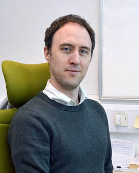 Mark Humphries Mark Humphries The OHBM is dedicated to understanding the anatomical and functional organization of the human brain using neuroimaging. But how to best use brain-activity measurements, including human neuroimaging, to understand computational mechanisms remains an open problem. “Mapping the brain does not by itself reveal the brain’s computational mechanisms” says Niko Kriegeskorte, past chair of the OHBM Communications Committee. “Therefore one of the strategic priorities in the OHBM Communications Committee has been to explore the interaction between computational neuroscience & human neuroimaging.”
Here, we had the chance to discuss the current state and future of computational neuroscience with Mark Humphries, senior research fellow at the University of Manchester, Chair of Computational Neuroscience at the University of Nottingham, and talented blogger. We found out about research environments in different countries, mindful language use in neuroscience, Mark’s outlook on the future of network neuroscience, and his top three tips for those starting out in computational neuroscience. By Elizabeth DuPre and Kirstie Whitaker The open neuroimaging community is great and growing every day. This month saw the first of a series of Open Science Demo Calls. Brought to you by the OHBM Open Science Special Interest Group, these live streamed calls are a chance to hear from the developers of open neuroimaging tools. We'll use these calls to build connections between all members of the OHBM Open Science community and to tell the stories of the people making outstanding and reproducible neuroscience happen. 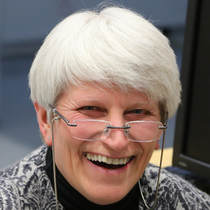 Aina Puce (photo by Ritta Hari, @aivoAALTO) Aina Puce (photo by Ritta Hari, @aivoAALTO) Professor Aina Puce is the Eleanor Cox Riggs Professor in the department of Psychological and Brain Sciences at Indiana University, Bloomington, and a senior editor at Neuroimage. She has followed a career path that is now becoming more common in human brain mapping, starting firmly rooted in the methods end but, over time, gradually shifting focus towards understanding complex patterns of behaviour. To do this, she has made use of a number of imaging techniques, exploring ways to extract converging lines of evidence.
Here, we find out how her interests changed throughout her research, the promises and pitfalls of multi-modal imaging, and why you should not be discouraged by rejections but instead focus on and be motivated by the paper acceptances and other highlights in your career. |
BLOG HOME
Archives
January 2024
|
 RSS Feed