|
by Athina Tzovara, Julia Kam, Valentina Borghesani, AmanPreet Badhwar ‘If you never did you should. These things are fun and fun is good’ - Dr. Seuss
Live Review for Kids The OHBM Diversity and Inclusivity Committee is exploring an exciting and new direction this year: we will be engaging kids in the scientific review process! We asked five prominent scientists in the field of brain mapping and neuroscience to write a short article explaining their research to kids. The articles are written for the Frontiers for Young Minds (https://kids.frontiersin.org/, a journal dedicated to young readers of 8-15 years old. Once written, the articles are assigned to five young reviewers, who will work together with a neuroscientist mentor to critique the articles and prepare questions for the scientists.
0 Comments
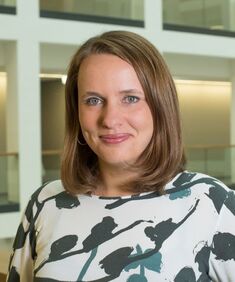 Claudia Buss Claudia Buss by Ekaterina Dobryakova
In preparation for this year’s Annual Meeting, we spoke to one of the keynote speakers, Dr. Claudia Buss. Claudia is an Associate Professor at the University of California, Irvine and a Professor of Medical Psychology at the Charité University Medicine in Berlin. In a virtual meeting, I sat down with Dr. Buss to discuss her captivating research in the field of developmental programming and newborn infant neuroimaging. Ekatarina Dobryakova (ED): Dr. Buss, thank you for dedicating your time for this interview. Before we get into more specific questions, I was wondering whether you mind sharing a bit about how you came to do the work that you're doing, and what got you to follow this passion in research. Authors: Claude Bajada, Nils Muhlert, Ilona Lipp
Infographic: Roselyne Chauvin Expert editors: Alfred Anwander, Jurgen Gatt Newbie editor: Caroline Jantzen Introduction Neuroanatomy is one of the most exciting topics in neuroscience! Some readers may disagree, but for now, humor us and read along. With the help of this On-Demand post, we will convince you not only that anatomy is a useful endeavour but that it is one where much beauty is found. Our journey starts with the fundamental notion that the structure and the function of objects are tightly coupled; sometimes in ways that are not obvious. Understanding the complexity of the brain’s structure, hopefully, allows researchers to build more accurate models of brain function. |
BLOG HOME
Archives
January 2024
|
 RSS Feed