Search the site...

organization for human brain mapping
  • BLOG
  • Tutorials
  • Media
  • Contributors
  • OHBM WEBSITE
  • BLOG
  • Tutorials
  • Media
  • Contributors
  • OHBM WEBSITE

 BRAIN MAPPING


Q&A with Lara Boyd: Turbo-launching OHBM 2017 and future of translational stroke imaging

8/30/2017

0 Comments

 
By David Mehler

​The Local Organising Committee (LOC) at OHBM 2017 achieved a remarkable feat. With public health experts voicing concern about the spread of the Zika virus from South to Central America, it was decided that it was too risky to expose so many young OHBM members to potential infections in Puerto Rico, this year’s original site for OHBM. At this point, the Vancouver LOC stepped forward. They organised an entire, major international neuroimaging conference not in four years, as planned, but in one.

Here we speak to Lara Boyd, Professor of translational neuroscience at the University of British Columbia (UBC),  chair of the Vancouver LOC, and TEDx sensation. We find out about the challenges of setting up OHBM at such short notice, and about her work mapping out rehabilitative medicine in stroke survivors:
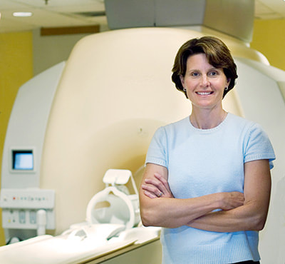
Picture
Lara Boyd
David Mehler (DM): It is my great pleasure to introduce Lara Boyd – she’s a professor here at UBC, where she leads the Brain & Behaviour Lab. Lara, perhaps you could give us some insight into chairing the organization of the 2017 OHBM meeting?
Lara Boyd (LB): It was an exciting year to chair. In case you missed it, we were supposed to be in Puerto Rico in 2017, and in Vancouver in 2020. Because of circumstances related to world health, we moved it up somewhat suddenly. It only came together through everyone scrambling and working really well together. We were lucky that the venue was available and we were available on short notice. The most fun we had was putting together the local organizing committee symposia, where we could showcase some of the science we do here in British Columbia. That was the best part!

DM: Can you tell us what it takes to host OHBM?
LB: First, we worked with the organizing committee of the OHBM to find the venue, and select the different speakers for all the symposia; that was a fun process that we really learned from.  We worked with the convention centre group just on the physical location; that was less fun but still exciting – particularly in a place that looks like this [looks out the window]. After that, we worked with the student group to make sure we had the venues and the social events planned and they had spaces for Brain Me Out, the Hackathon, those kind of things. Last, we got to put together the symposia. Now, at this point, when we’re all here, we get to just sit back and enjoy and show off a little bit, and that’s been the most fun part.

DM: There have been many highlights at the conference so far; for instance, Tal Yarkoni’s input on the statistical implications of fMRI analysis, and then a session on myelin imaging at which you were co-chair. What were your personal highlights of the conference?
LB: I loved Tal’s talk --- and I’m not a statistician but he just made that info so accessible. My lab is excited to go home on Monday and try it and see what happens. I also loved the Talairach Lecture, I think Carla Shatz did such a nice job and it’s something I knew nothing about – not my field, not my expertise – so to just sit back and watch how science in one area progressed to something totally unexpected, and she was able to take that knowledge and translate that into something that’s really going to help people with Alzheimer’s and other dementias, that was really exciting. It was just a wonderfully put-together talk.

DM: You mentioned translation – your lab is heavily involved in translational neuroscience, particularly in stroke rehabilitation. Your work has contributed to our understanding of how therapies in stroke can work. You’ve more recently shown plasticity even within myelin - very exciting work! It’d be interesting to know what got you into stroke research and what you find particularly interesting about this field.
LB: I actually started my professional career as a physical therapist. It didn’t last that long – only about a year. Part of that was because my stroke patients just didn’t get better. I had that sense that I was a car mechanic and didn’t understand how the engine worked. So I went back to school to become a neuroscientist to understand how the brain worked in the hope to translate that information back into therapies for stroke. That’s what led me into the field and it was perhaps good timing, as that was right when the field took off.

I believe the first OHBM was in 1995 and that’s when I started my doctorate, so I just grew up with the meeting, and with the field in general. It’s just been a set of really lucky circumstances that has allowed our science and translation to advance so rapidly.

I’ve always said that I’m on the consumer end of the neuroimaging spectrum. We take these beautiful approaches that our physicists are designing and we use them to try to really unpack the changes that occur in the human brain. That’s why my lab is called the “Brain and Behavior Lab”. We also try to find out what behavior enables those changes and try to map them. We try to take that information and leverage it into therapies for people with stroke and try to really speed their recovery. We try to enable greater recovery than we’re currently seeing by improving our basic understanding of how the brain changes.

DM: At this conference we witnessed that the field of translational neuroscience is rapidly growing. For many young researchers with clinical backgrounds that want to pursue a career in neuroimaging, what would be your top three tips for starting out?
LB: First, no question is a dumb question. Don’t be afraid to go up to a senior scientist, or just to someone in an area that you’re unfamiliar with or comfortable with and ask that question. Ask about the field – how did they get into it? What kind of things led them to that? We need to remember that we were all junior scientists and just starting out at one point. I find that everyone is really happy and helpful in sharing their knowledge.

Second, make as many connections as possible. You can see the field is incredibly diverse. There are many different imaging platforms. The future I’m seeing is where there is going to be much more multimodal imaging. We can’t be an expert in all of those areas, so we’ll need a lot of good friends. We can start to translate information from different findings in different research studies to understand this marvelously complex thing, the brain.

​
Last, build connections with your peers. These are the colleagues that are going to be reviewing your grants and your papers and in the future these are the people that are going to give you students for your lab as you move along. The more interconnected you can become with your peer group as you rise up through the ranks, the better suited you’ll be when you need that friend who knows a technique when you don’t. You can call upon them and they can really enrich your science.
DM: Thanks Lara. Last, can you give us a bit of an outlook for the field of translational neuroimaging for stroke rehabilitation and where you see this field going within OHBM?
LB: In stroke rehab right now we’re actually a little bit stuck. Lately, we’ve had a bunch of clinical trials that failed. They failed to show any benefit beyond regular care. In part I think it’s because we treat stroke as if it were a single condition - any of us who have seen stroke patients know that they’re marvelously different.

So what we’ve become really interested in, in my group, is understanding biomarkers that can help us sub-categorise people with stroke. We then use those biomarkers to predict what recovery patterns we might see and which treatments are going to be best for which patients. Our stroke recovery biomarkers are all neuroimaging derived. So we can take a human stroke patient, we can use maybe diffusion or myelin water imaging to understand the residual brain structure, understand how that patient may be compensating through different networks in functional patterns, how their cortical excitability is changing with Transcranial Magnetic Stimulation. Then we start to build algorithms and models that take each of those pieces of information and put them together to build a more complete portrait of that patient. We then use that information to predict what may be the best therapy for them.

I really think that as we become better consumers of these many different multi-modal types of imaging we can really put them together in a meaningful way. That’s what will move stroke rehabilitation forward, as it will allow us to understand that unique complexity of each patient.

It’s sort of what you might think of cancer treatment: cancer treatments are very personalized, highly tailored to each individual. We want to use neuroimaging to do the same thing with our stroke patients. That’s the future I envision and I hope we’re moving rapidly towards it.
​
Thanks Lara, and many thanks to Sarabeth Fox for filming.
0 Comments

OHBM 2017 Abstract Highlight: Neuroplasticity Associated with Reduced Depressive Symptoms after Cognitive Training for TBI

8/16/2017

0 Comments


Q&A with Dr Kihwan Han
BY EKATERINA DOBRYAKOVA ​

Depression is a common psychiatric disorder amongst individuals with traumatic brain injury (TBI). Up to 77% of individuals with TBI have been found to experience depression. What can brain mapping tell us about depression after TBI? I met with Kihwan Han from the Center for Brain Health at the University of Texas at Dallas to talk about his recent research: an eight-week intervention that aimed at battling depression symptom severity in individuals with TBI.
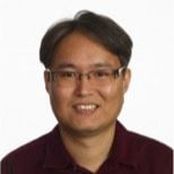
Picture
Dr Kihwan Han
Ekaterina Dobryakova (ED): First, can you tell us about the findings of this particular research project?
Kihwan Han (KH): In this study we investigated whether 8 weeks of cognitive training would reduce symptoms and severity of depression in individuals with chronic TBI. Indeed, we found reductions in depressive symptoms in individuals with mild-to-severe depressive symptoms compared to individuals with minimal depressive symptoms. Decreases in depression severity were also associated with improvements in self-reported post-traumatic stress disorder (PTSD) symptom severity, TBI symptom awareness and functional status. Further, reduced depressive symptoms were related to thickening of regional cortical gray matter and reductions in abnormal brain connectivity (Figure 2).
Picture
Figure 2. The pattern of right dorsal prefrontal connectivity in association with reduced depressive symptoms after cognitive training in individuals with TBI. The right dorsal prefrontal cortex also exhibited changes in cortical thickness modulated by reduced depressive symptoms after training.

ED: How long have you been involved in TBI research and why are you interested in TBI?

​KH: I started doing TBI research in 2011 as a post-doc in the neurology department at Washington University in St. Louis. The lab was exploring all aspects of TBI, including animal models.

ED: Do you think animal models of TBI, such as controlled cortical injury, can be informative?
KH: There is definitely strength in TBI animal research since a lot of variables can be controlled, with the ultimate goal of looking at what happens to the brain of humans when a TBI is sustained. The issue with animal research is that we are interested in how the brain functions after TBI but the animals and humans are different on the functional level. For example, the symptoms of TBI in humans are more complex.

ED: As you were just saying, TBI is a complex issue and usually researchers study aspects of different TBI severities. At OHBM, your presentation addressed not only TBI but also a complex mental disorder, specifically depression in individuals with TBI. What do you think is the value of studying psychiatric symptoms in clinical populations?
KH: TBI is a complex neurological event that leads to a variety of comorbidities. In military personnel with TBI, many researchers are looking at PTSD. But actually, depression is one of the most common psychiatric disorders in individuals with TBI. I think more and more researchers are starting to look at the different consequences of TBI, and depression should definitely be one of them, in my opinion.

ED: What do you think is the appeal of your work for broader audiences?

KH: I hope my research appeals to individuals with TBI and their loved ones who strive to get information about depression in TBI. My goal is for my research to give hope that there are rehabilitative solutions for individuals with TBI who also have depression. At the same time, I think my work should also appeal to scientists outside of the cognitive and clinical neuroscience fields, such as engineering. From the engineering point of view, TBI is a complex problem: one has to extract meaningful data from such a heterogeneous group of subjects. My background is actually in engineering and I started my postdoc with neurologists because they really needed help in analyzing the data, and were looking for someone who can bring a different perspective to the data. More people with diverse educational backgrounds are starting to do TBI research, so I think my research is appealing to people of different backgrounds.

ED: Can you briefly describe the next steps for your project?
KH: In the current project, we simply looked at the correlation between thickening of the gray matter and reduction of depressive symptomatology after an eight-week intervention. The next step will be to try and apply neuromodulation techniques to brain regions that were associated with a reduction of depressive symptomatology. Maybe neuromodulation can also lead to the reduction of depression symptoms.
0 Comments

OHBM 2017 Abstract Highlight: MULTIDIMENSIONAL MRI SUBTYPING OF AUTISM SPECTRUM DISORDERS

8/4/2017

0 Comments


Q&A WITH HONG SEOK-JUN

By Tzipi Horowitz-Kraus

This interview series highlights abstracts from the OHBM meeting that were identified by the Program Committee as “potentially newsworthy”. Abstract authors were asked to explain their research in more detail, discuss the context of their findings, and the possible implications for the field.
Autism spectrum disorder (ASD), is a developmental disorder that contains extensive symptomatic variability across people. There are presently no biomarkers to guide diagnosis of ASD - it continues to be diagnosed only after a child does not demonstrate the appropriate communication skills. In order to work toward identifying potential biomarkers from brain data, Hong Seok-Jun, a student with Prof Boris Bernhardt from the Montreal Neurological Institute at McGill University in Canada focused on Multidimensional MRI subtyping of ASD in adults. I met with Hong Seok-Jun to better understand his motivation to conduct this study and the implications of his findings.
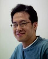
Picture
Hong Seok-Jun
Tzipi Horowitz-Kraus (THK): If you had to explain your study to your non-brain mapping friends, what would you say?
Hong Seok-Jun (HSJ): Autism spectrum disorder is a common neurodevelopmental diagnosis. Previous research suggests that there is likely no single autism type, but rather a collection of different subtypes of autism that may each be associated with different biological causes and behavioral symptoms. In our project, we built a novel approach to subgroup a larger population with a diagnosis of autism based on multiple MRI measures of cortical morphology. We could indeed identify multiple subgroups with different imaging features and functional network anomalies. Using a machine learning framework, we furthermore show that information about subtype was useful in predicting the severity of behavioral symptoms in individual cases, possibly suggesting clinical utility of the proposed framework.

THK: What motivated you to go into your particular area of research?
HSJ:
In autism, previous imaging studies have reported mixed findings, failing to converge onto a common pattern of brain anomalies. This may challenge current diagnostic procedures and impede person-specific therapy. We hypothesized that a main source of the inconsistency across previous findings may relate to heterogeneity across the autism spectrum itself. Our study presented at this OHBM conference addressed the heterogeneity of structural brain phenotypes as well as their potential implication in behavior and brain function in autism spectrum.

THK: Why do you think your abstract was selected as newsworthy?
HSJ:
Our study addresses a timely question in the field - namely the identification of different subtypes within a larger and rather diverse clinical population. By integrating advanced MRI features of vertical and horizontal cortical organization (such as cortical thickness, cortical interface blurring, surface area, and geodesic distance), our framework may offer a biologically meaningful way to group subtypes of autism phenotypes which are characterized by specific brain morphology and behavioral symptoms. Finally, our study makes use of both unsupervised as well as supervised learning techniques to address these high dimensional datasets, and can thereby derive predictors of symptom severity at the individual subject level.

THK:  What are your next (research) steps going to be, given these findings?
HSJ: In future studies, MRI subtyping approaches could be complemented by other neuroimaging modalities, more extensive behavioral assessments, as well as genetic information.  This would collectively result in a comprehensive and multidimensional characterization of the autism spectrum. Furthermore, our work may provide a useful framework to address the heterogeneity often found in other neurodevelopmental disorders. 
Picture
Our novel imaging framework allows for an objective identification of multiple biotypes in the population of autism spectrum disorder (ASD), based on advanced MRI markers for cortical morphology. The discovered ASD subtypes indeed reveal distinctive neuroanatomical phenotypes, suggesting highly heterogeneous biological substrates across the autism spectrum.
0 Comments

OHBM 2017 Highlights: the Inaugural Chinese Young Scholars Meeting

8/2/2017

0 Comments

 
BY DENGFENG HUANG

Travel makes you richer --- it lets you experience new landscapes, languages and ways of thinking. This philosophy sits well with OHBM. Our organisation promotes travel and discussion amongst brain mappers from different corners of the globe and different sections of society.

As part of this cultural engagement, we currently have 5 chapters, representing brain mappers in the Alpine region, Turkey, China, Korea and Latin America. These Chapters allow researchers from clear geographical areas to pool their knowledge and resources, and speak with a unified voice both within OHBM and to stakeholders in science more generally. An excellent example of the knowledge that can be gained - not just within these groups but by outside observers - can be seen from the first meeting of Chinese Young Scholars.

This inaugural event was co-organised by
Chao-Gan Yan, and originally covered by Dengfeng Huang in Mandarin on WeChat. Here we present an English translation, revealing to those less adept at reading hànzì just how the event went, and offering useful career advice for early career researchers around the world.


The first Annual Event of Chinese Young Scholars was successfully held on June 27, 2017 during the OHBM Annual Meeting in Vancouver, Canada. Recent years have witnessed a rapid development of brain research, ranging from molecular mechanisms to neuroimaging and computational modeling. This surge of brain science research has brought together scientists from a variety of backgrounds, such as neuroscience, psychology, physics, statistics, and engineering. Interdisciplinary communication and collaboration have hence become increasingly important. In addition, it has become increasingly clear that young scientists often experience confusion and bottlenecks in their careers. With these thoughts in mind, we hoped to provide a platform where: 1) Chinese researchers can communicate, discuss, and collaborate on cutting-edge neuroscience topics and methods, and 2) young scholars can seek advice from senior researchers on the route to a successful scientific career.

​The 2017 Annual Event of Chinese Young Scholars for Human Brain Mapping received support from the OHBM China Chapter and the OHBM Communications Committee. After the event was announced in May, it attracted enthusiastic responses worldwide. We received registrations from over 130 Chinese researchers based in over 10 countries and regions, including Mainland China, Hong Kong, Taiwan, the United States, Germany, Canada, Australia, the United Kingdom, South Korea, Singapore, and Switzerland. Our attendants ranged from graduate students, junior researchers, senior researchers, professors, to colleagues from industry.​

The event commenced with the host, Yuhua Guo, introducing the organizing committee: Chaogan Yan, Yuan Zhou, Yina Ma, Dengfeng Huang, Yuhua Guo, Bin Lu, Shengchuang Feng, Chunliang Feng, Yinshan Wang and Qing Wang.
Picture
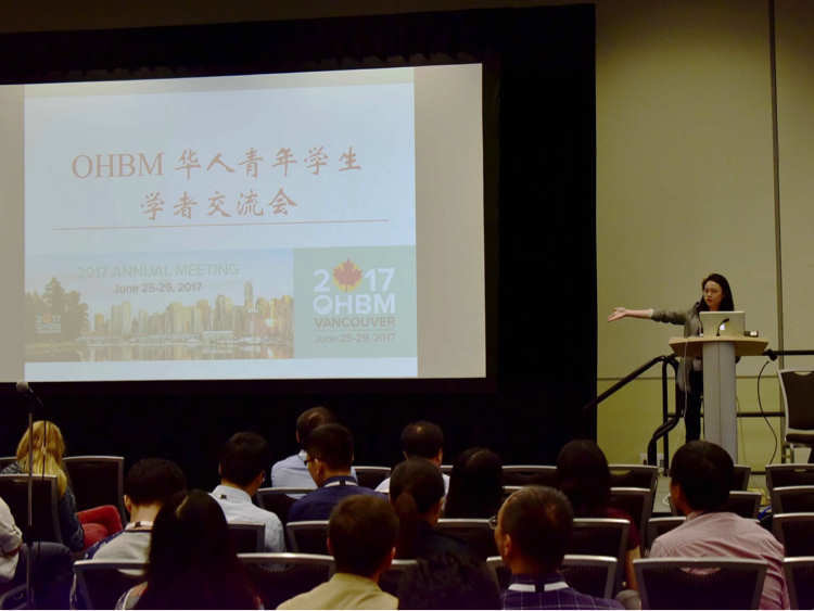
The host, Yuhua Guo, introducing the organizing committee.
Picture
Professor Chaogan Yan, from the Chinese Academy of Sciences, was the mastermind behind the event. Although Professor Yan was unfortunately unable to attend, he recorded a video to introduce the rationale for launching this event and to wish it success.

​The formal schedule started with two keynote speakers sharing insights into developing research methods and choosing research questions. The first speaker was
Professor Yihong Yang, Chief of Section of Magnetic Resonance Imaging and Spectroscopy and Senior Investigator from the National Institute of Drug Abuse, USA. Through sharing his latest research, the Role of Functional Magnetic Images in the Study of Psychiatric Disorders, Professor Yang inspired young scholars to tackle scientific questions with rigorous thinking, careful experimenting, and dedication to seeing things through.


The second speaker was Professor Tianzi Jiang, director of the Brainnetome Center at the Chinese Academy of Sciences. Professor Jiang shared his contribution on the brainnetome atlas, an in vivo map with fine-grained functional brain subregions and detailed anatomical and functional connection patterns for each area, and the application of this brainnetome atlas to neuroscience research. Professor Jiang emphasized the importance of choosing a meaningful research direction and being persistent in pursuing it.
Picture
Prof Yihong Yang discusses the use of fMRI in studying psychiatric disorders.
Picture
Prof Tianzi Jiang on the Human Brainnetome Atlas.

​Professor Yihong Yang provided advice on how to be a successful research student, based on his experience back in the 80s. First, he suggested that you should always try to turn adversity to your favor, whether the adversity is from interpersonal relations, developing theories, or interpreting results. Professor Yang advised that if you ever get stuck, it helps to divert some attention to other useful tasks, such as learning new knowledge or skills. This could not only help reduce anxiety, but also build some sense of achievement. Second, when comparing Chinese students with western students, Professor Yang pointed out that although Chinese students are generally diligent, they sometimes lack creativity and independence. He encouraged young scholars to cultivate their own ideas in addition to working hard, to better prepare them for becoming an independent researcher.


​Professor Tianzi Jiang shared insights on identifying research interests. In his career, Professor Jiang switched research interests multiple times and sought opportunities both in China and abroad. He advised that people could only find their passion once they understood themselves. Importantly, when searching for the ideal research topic, one should not only focus on the data at hand, but also envision important research trends in the long run based on existing knowledge. It would be best if one can predict research directions in the next decades and work towards those directions.

Professor Jing Sui shared her experience of switching from engineering to neuroimaging, emphasizing the importance of self-motivation. She stressed that young scientists should be more motivated and proactive in their projects, and not just be satisfied with what their supervisors instructed them to do. She echoed Professor Yang’s advice that becoming an independent thinker is a crucial first step to a successful career in science.

Professor Yina Ma added that young scholars should "fully estimate and prepare for the difficulties you may encounter in an academic career ... when you see researchers being successful, it must be that they have already overcome countless frustrations and difficulties. If you are still willing to carry on with research after considering all the possible pitfalls (such as having your manuscript rejected, or having to change your research topic), then a scientific career may be truly for you!”

Towards the end of the session, Professor Chun-Yi Lo reminded early career scientists to cherish the time they have now. “It’s your best time to fully concentrate on a particular question, while having the chance to embrace and prepare for all sorts of possibilities in the future. Do value it and make the most out of it!"
Picture
The audience thanked the panelists with a huge round of applause. We took group pictures to conclude this inspiring and memorable event.

​After the meeting, we enjoyed a group dinner and live music at Malone's Social Lounge and Tap House. Young and senior scientists mingled in a more relaxed atmosphere, discussing science and research. We were pleased to receive lots of positive feedback from the attendants. One remarked that “[The event] is simply beyond my expectations! It is really amazing to discuss frontier neuroscience research topics with senior investigators, to get advice for career development, and to know other people’s research interests and form collaborations." The success of the first Annual Event of Chinese Young Scholars for Human Brain Mapping was highlighted at the closing ceremony of OHBM 2017. We sincerely look forward to meeting everyone again next year in Seoul, South Korea!
0 Comments

    BLOG HOME

    ​TUTORIALS

    ​MEDIA

    ​contributors

    ​OHBM WEBSITE


    ​

    OHBM OnDemand 
    ​Education Platform


    RSS Feed

      blog survey

    Submit

    Archives

    February 2021
    January 2021
    December 2020
    November 2020
    October 2020
    September 2020
    June 2020
    May 2020
    April 2020
    March 2020
    February 2020
    January 2020
    December 2019
    November 2019
    October 2019
    September 2019
    August 2019
    July 2019
    June 2019
    May 2019
    April 2019
    March 2019
    February 2019
    January 2019
    December 2018
    November 2018
    October 2018
    August 2018
    July 2018
    June 2018
    May 2018
    April 2018
    March 2018
    February 2018
    January 2018
    December 2017
    November 2017
    October 2017
    September 2017
    August 2017
    July 2017
    June 2017
    May 2017
    April 2017
    March 2017
    February 2017
    January 2017
    December 2016
    November 2016
    October 2016
    September 2016
    August 2016
    July 2016
    June 2016
    May 2016
    April 2016

stay connected with ohbm!


become a member

Telephone

952-646-2029

Email ohbm

EMAIL BLOG TEAM
Header image created by Thiebaut de Schotten & Batrancourt  
www.brainconnectivitybehaviour.eu