BY NIKOLA STIKOV At the OHBM 2015 Annual Meeting in Honolulu, HI, the OHBM Communications Committee (or ComCom as we like to call it) was created by the OHBM Council. They appointed Randy Gollub as its first Chair and Niko Kriegeskorte as Chair-Elect. Over the fall and winter Randy and Niko worked with OHBM staff to recruit volunteers and create a structure for this new and exciting initiative. The first step after assembling a group of eager and extremely talented Committee members was to divide the large group into four different specialized teams and a quarter of the members were assigned to the ‘Member Communication’ team. Our task was daunting. How do you communicate year-round with a membership that spans so many disciplines, continents and backgrounds? Do you create separate newsletters, Facebook pages and YouTube channels? Do you hold monthly meetings in which you assign specific tasks targeting specific audiences? Or do you put all this under one umbrella, call it a blog, and let the OHBM members decide what to do with it? If you are reading this, then the answer is obvious. We began with a blog because it was the most inclusive (and easiest!) platform we could think of, and then allowed everything else to evolve organically. The “Member Communication” Team became the “Blog Team” and over the past year we have published 51 blog posts, featuring over 40 contributors from 11 countries. The blog has received over 165,000 page visits from 63,000 unique visitors, we’ve created 12 videos for our YouTube channel and OHBM had over 345,000 impressions on Twitter. The OHBM Communication Committee’s work has also been featured in the Huffington Post and cited by the New York Times. Not bad for a one-year old... None of this would have been possible without the vision and guidance of Randy and Niko, the support and resourcefulness of the ComCom team captains Jeanette Mumford and Lisa Nickerson (Website Team Captains 2016/2017), Cyril Pernet (Social Media Team Captain) and Kevin Weiner (Media Team Captain), and the organization and professionalism of our Communications Manager, Stephanie McGuire. Finally, there is our core blog team that spans five time zones that are 15 hours apart, yet somehow manages to stay awake during grueling monthly discussions about guest posts, calendars, spreadsheets, SOPs and Trello cards, only to go back to their computers to do more pro bono work than they ever volunteered for. The fact that they keep coming back, month after month, means that we have found a way to have more fun than this post makes it seem. Don’t believe us? We have a suggestion for you-- become a contributor!
As we are transitioning from baby to toddler, we are looking for friends and role models to look up to. And while one-year olds are known to stumble now and then, they are also endlessly curious and growing at a rapid rate. We hope to continue to grow, evolve, and become more steady on our feet as we figure things out-- and most of all to be receptive and responsive to our community in the years to come!
0 Comments
![]() BY GUEST AUTHOR ERIKA RAVEN This post originally appeared on the ISMRM blog and in the MRM Highlights magazine. Republished (and slightly modified) with permission. Karla Miller is a professor of biomedical engineering at the Oxford Center for Functional MRI of the Brain (FMRIB, pronounced “fim-rib” for short). She directs the FMRIB Neuroscience Physics group, which specializes in many projects, from pulse sequence development to biophysical tissue modeling. More recently, she’s been a key figure of the UK Biobank, a mega-sized data initiative charged with imaging 100,000 adults by 2022. Karla is also a plenary speaker at the upcoming OHBM meeting in Vancouver, education chair of this year’s ISMRM meeting in Honolulu, and is poised to chair the entire ISMRM program for the 2018 meeting in Paris. In our interview, Karla makes connections between the many themes in her life, which ultimately are resolved by finding the right balance. Erika Raven (ER): You’re one of the few people that feel comfortable straddling the line between ISMRM and OHBM. Do you see a synergy between these two societies, or would you rather they keep running on parallel tracks? Karla MIller (KM): I think it’s incredibly important that people who are developing MRI techniques don’t do so in a vacuum. I’ve benefited tremendously from being at the FMRIB center. Although I’m in a physics group, I rub elbows with people on the analysis and neuroscience side. I think it’s important for people who are developing these sequences to understand how neuroscientists will want to use them. Cross society outreach is something I am keen to do as part of becoming chair of the ISMRM’s annual meeting program committee (AMPC) in about 6 months. ER: How did you first become involved with ISMRM and what led you to become this year’s education chair? KM: I first attended the ISMRM in Philadelphia (1999) and I have attended every ISMRM since. One of the first official roles I held was to serve on the AMPC. The AMPC is the hardest working, but also the most exciting, committee to be a part of. Now for this year’s ISMRM, I am coordinating the education for Hawaii, and then at the Paris meeting in 2018 I’ll be chairing the entire program. I’m incredibly grateful to Dan Sodickson for appointing me - although as the huge task ahead really hits me, I might save my thanks until the meeting is a wrap! ER: You’ve given many educational seminars. What is it about MRI education that you like? KM: I absolutely love teaching. Beyond it being immensely satisfying to help people grasp difficult concepts, I think it's a good experience for the lecturer to think hard about the material. It’s an interesting challenge - can I do a better job of teaching this to other people than it was taught to me? ER: Your work is multifaceted - can you explain your primary research themes and how those came to be? KM: My training was very much in pulse sequences and image reconstruction. And so I still have a big chunk of my group working in that area. In the past few years, I’ve become interested in the idea that we can improve our acquisitions and reconstruction by taking a lead from how people analyze their data. We tend to think of this as a linear process – you try to get the best data you can and then you analyze it. But there are tricks that we can learn based on how people analyze the data that would enable us to improve the acquisition and reconstruction itself.
ER: I would imagine he would be useful! You also study biophysical modeling and ex vivo imaging of tissue microstructure. Can you tell us about that? KM: We’re acquiring microscopy data so we can close the loop between what is the biophysical model, what is the MRI data, and what is the actual measurable microstructure. The key aspect of our experiments are that we have all three things –MRI and microscopy in the same tissue samples, and a proposed model linking them. By actually having a measurement of the underlying microstructure, it guarantees thatis if you’ve got your model wrong, you are the absolute first person who is going to know. Not just, “can I take a biophysical model and show that it kind of matches the data”, but “can I actually take something that I know reflects the underlying microstructure, make a prediction through some biophysical model, then say - YES - that is exactly the MRI signal that I measured”. And it’s a really hard thing to do. ER: That was like a mission statement! KM: Putting this process to work, we’ve been looking at diffusion based estimates of fiber dispersion. We use microscopy techniques to essentially ask what aspects of the microstructure you need to incorporate to accurately predict what the diffusion signal looks like. It’s a project that has a true palpable output, and interestingly it’s created a signature that we hadn’t expected to find. We’ve now demonstrated that this particular effect also exists in the Biobank data - so it’s a real effect, which is potentially a signature of something biologically interesting. More importantly, we’ve managed to have a first go at what it might look like to actually close the loop of biophysical modeling, microscopy, and MRI acquisition. ER: I really like that turn of phrase, closing the loop. And since you mentioned the UK Biobank, I’ve given myself permission to bombard you with Biobank questions! To start, when did you first become involved? KM: What I’m actually doing right now as you’re asking me this question is looking in my emails to see when I had my first Biobank email logged. 2008! Email from Paul Mathews, basically asking if we would be interested in getting involved in the Biobank. It’s quite a project – scanning 100,000 subjects. And although there is quite a long author list on the paper that we published this year, that doesn’t even begin to cover the number of academics involved, let alone the enormous staff that is entirely dedicated to the project. As one colleague said – its behemoth. In a good way. ER: What do you think will change from having 10,000 scans to 100,000 scans? KM: One of the most exciting aspects of Biobank is that it’s an entirely prospective study: it has no particular disease focus, but is playing the numbers. Most of the participants in this huge cohort have yet to show symptoms of major disease, but we’ll be able to follow their health records as that changes. So, for example, we expect 2000 new diagnoses of Alzheimer’s and 50 new diagnoses of ALS over the next five years from participants who were pre-symptomatic at the time of imaging. The value in Alzheimer’s is obvious, but for rare diseases like ALS, that is a needle in a haystack. You just can’t find those subjects otherwise. It certainly might provide you with markers for tracking response to therapy or disease progression.
ER: It sounds like the translational aspect of this research might become even more important now, such as borrowing techniques from other fields that have already been established and validated for big data sets.
KM: It’s partly techniques and it’s partly culture. The same thing with open science – I know it’s the right thing to do, but there is part of me that thinks, “Ahh!, it’s going to be yet another thing I have to adhere to”. But once we have a culture of doing it, everybody looks back and says, “What were we thinking?” ER: A more general question, what brought you to the academic life? Did you have any major influences that led you down this path? KM: I got very interested in the brain when I was a kid. My mother had to have pretty drastic brain surgery when I was about 12 or 13. It really struck me - the idea that it might fundamentally change who she was. When I went to university, I started out as a psychology major. I was taking a cognitive psychology class in maybe 1995 when I saw functional MRI in a textbook - totally state-of-the-art . I was so impressed with what it had to offer compared to current methods for studying cognition. I also thought maybe the way I could have an impact was to develop the technology and move towards the engineering side. And so it’s kind of nice for me now that I’ve done the engineering side in anger for about 10 years, and I’m able to shift towards getting back to neuroscience. And for me that’s incredibly rewarding. ER: It’s like you’re closing your own personal loop. KM: There’s a theme there, isn’t there? ER: Now for some words of wisdom. What things did you learn along the way that you feel would be important for people who are just starting out? KM: I have to be profound on short notice! Well… Going into science with a great deal of passion, and a great love of what you’re doing is absolutely critical. Particularly if you want to stay in academia, because let’s face it - academia is a tough world to get by in. One of the things I did sort of instinctively early on was to look towards people who were a year or two ahead of me, doing the kind of science I wanted to do. If I have to name names – Brian Hargreaves and Bill Overall. They were my role models. I tried to see what it was that they were doing at my stage to get where they were. That sounds simplistic, but honestly, that was what I did. And it’s good advice. ER: It has come up repeatedly that you frequently go outside your comfort zone. That’s sometimes a scary thing to do – what drives you to change? KM: You know how I would sum this up… For me - and I know this is not true for everyone - being an expert is boring. To some degree, the fact that I’m the one “blah blah’ing” in this interview the whole time, from my perspective, it’s flattering but not stimulating. It would be far more fascinating for me to be asking you about what you’re doing and learning about what you’re doing. Being an expert is, for me, it’s the way you earn the opportunity to be an inexpert - that is the fun bit. That said, I wouldn’t encourage people to just jump from one thing to the next willy nilly, because you’ll never become an expert in anything, and that’s also not good. You have nothing then to leverage. So you have some safe stuff, and some risky stuff, and you’re hopefully pushing your personal envelope the whole time. It’s kind of about finding the right balance. SHRUTI GOPAL VIJ COBIDAS: OHBM Committee on Best Practice in Data Analysis and Sharing Neuroimaging researchers study both the structural and functional organization of the brain in health as well as a variety of neuropsychological conditions. Extensive exploration has been conducted over the past few decades using novel experimental and analysis techniques. However, recent evaluations of these techniques have highlighted concerns that published scientific results are less reliable or reproducible primarily due to the lack of transparency in research practices. To address the need for outlining principles of scientific research that will increase transparency and reproducibility in human neuroimaging, the Organization for Human Brain Mapping created the Committee on Best Practices in Data Analysis and Sharing (COBIDAS). COBIDAS has initiated efforts in distilling best practices for open science in human neuroimaging and has compiled suggestions for specific research practices to support open data and open methodology. These suggestions were composed in a report that was drafted and ratified using community collaboration. While the report itself provides a detailed description of best practices as well as approaches to avoid, a commentary that reviewed the impact of COBIDAS and the challenges ahead was published in February 2017. The commentary underlines the importance of reproducibility and highlights the different aspects of replicating a study including generalizability over methods, materials and results as shown in Figure 1. One practice that was universally recommended is the transparent and complete reporting of all facets of a study, allowing the critical reader to evaluate the work and fully understand its strengths and limitations. Additionally, thorough reporting will equip the reader/other researchers with a detailed knowledge of how to fully replicate the study. COBIDAS MRI report presents reporting checklists in Appendix D that researchers can follow while preparing manuscripts. These checklists are highly comprehensive and run the gamut of stages a human neuroimaging study passes through. The report also pinpoints specific bad practices, especially in statistical modeling and inference (Pages 11 & 12), and provides concrete recommendations for avoiding them. COBIDAS also suggest that researchers at all levels be mindful of these considerations while conducting individual studies and compiling manuscripts. Another strong recommendation from COBIDAS is data sharing as highlighted in section 7 (Pages 19-24). While sharing of results was strongly agreed upon by the imaging community and COBIDAS, some concerns were articulated by the community. The main concerns of the neuroimaging community outside of COBIDAS included data ownership, maintaining autonomy over results, concern of uncovering errors in analysis, cost of data sharing, and protection of subject privacy. COBIDAS posits that there are ways to overcome each of these concerns by not only being personally open to the fallible nature of individual research and welcoming collaborations initiated by data sharing, but also by emphasizing the role of the organization and the lab in making these practices widespread. COBIDAS also suggests that a culture of constructive criticism needs to be imbued in the community to allow researchers to work on reproducibility studies. They credit some studies such as Waskom et al, Whitaker et al and Pernet et al as well as the Montreal Neurological Institute that have become the flag bearers of open science by sharing their research data and analysis scripts. Such recommendations and practices are leading to the development of new data sharing methods and open science tools for neuroimaging. Some of the identified tools for data sharing at various experimental stages include the Brain Imaging Data Structure (BIDS); the CBRAIN web-based analysis service; the COINS service; the LONI pipeline; the Neurovault repository; and the FCP/INDI and ADNI data sharing repositories. The COBIDAS recommends using the many resources available to share data, analysis scripts, and well-written code, along with unthresholded result maps to allow precise meta-analysis and follow up studies. Finally, the COBIDAS acknowledges that while many of the practices suggested and recommended require individuals to change the way they approach implementation and reporting of a research study, advancement of these open science initiatives will only bear fruit by institutional support and direction. They posit that universities and research centers, as well as the journals themselves, should require data and code sharing. Foundations and grant agencies should also recognize and fund the explicit costs of data sharing. However, none of these will be able to single-handedly bring about the radical change that open science warrants. COBIDAS seeks a coordinated effort from individual researchers, research centers,universities,grant agencies, and it also invites professional organizations to accelerate the drive towards open science via extensive education and outreach. What does this mean for you and me as human brain mappers? We need to be more transparent in what we do. Not just because of the implications this has on clinical applications, but to make human brain mapping a more reliable field. Small changes in our approach to science such as documenting all the analytical methods applied while working through the projects and sharing this document on our personal sites or as supplementary material to manuscripts is a small initiative we can all take. Another simple task would be to share code written for projects on code sharing platforms such as github. At the level of the PI’s, making it a lab requirement to share this information with all the lab members including de-identified participant information and maintaining documentation for all projects: ongoing and completed would be speed up the process. This also makes it easy to follow up on projects once postdocs and graduate students have moved on. As they say- “Little drops of water make the mighty ocean”, if we all commit to do our part, we can make open science a reality and very soon. BY NILS MUHLERT Every year OHBM receives thousands of abstracts, each a snapshot of the intensive work of individuals or teams of scientists. Contained within this bumper crop of science are telling replication studies, novel experimental designs, methodological advances and fresh insight into the workings of the human brain. This harvest, whilst welcome, necessitates difficult decisions about which prize specimens to highlight as talks, and which to promote through posters. For those of us yet to work on program committees this process of selection can seem opaque - we can perhaps predict the established trends and exciting developments from year to year, but how do committees decide which emerging fields will pique the interests of the majority of OHBM attendees? Here we find out the decisions that were made in deciding on the OHBM 2017 program, through discussion with the Program Chair and Stanford Neurologist, Mike Greicius.
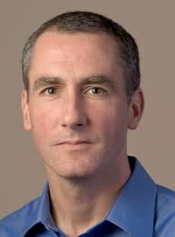
 ![]() Nils Muhlert (NM): First, can you tell us about your career path into Neurology and Neuroimaging? Mike Greicius (MG): I was a French major as an undergraduate but was able to get my pre-med classes done at the same time. I went to Columbia for medical school but was allowed to defer my matriculation for 1 year so that I could "teach English" in Prague and Paris, which, in fact, I sort of did. Taking a year off allowed me to hit medical school refreshed. It was as a second-year medical student at Columbia that I had my epiphany about neuroscience. I was in a lecture on aphasia and the professor, Richard Mayeux (now the chair of Neurology at Columbia), had a videotape showing a conversation with one of his Wernicke's aphasia patients. I was thunderstruck and knew, from that point, that I wanted to be a behavioral neurologist. In my head I summed it up as something like "why study the kidney or the liver when you can study the one organ that talks back to you?". I did my one-year of internal medicine at Columbia and then residency training in Neurology at Harvard, which is a fondly remembered blur. Fondly, mainly because it is blurry. At the time I was rather zombie-like, overworked, underpaid, etc. Those were three tough but formative years. In 2000, on finishing my residency I came to Stanford for an fMRI fellowship (with Allan Reiss and Vinod Menon) which I combined with a behavioral neurology fellowship (done at UCSF with Bruce Miller). The sun came out and everything changed. I was delighted to learn that my wife still loved me and that I could now spend time awake with my then 2-year old son. My big resting-state epiphany actually happened at my very first conference, which happened to be OHBM 2001 in Brighton. That was my first taste of resting-state fMRI. The Biswal paper had been published in 1995 but I had had no exposure to imaging research in residency. I recall sitting in my hotel's pub sponging insights from Dietmar Cordes, whose group at Madison was doing a lot of the early fundamental methods work in resting-state fMRI. I was really blown away by the power of this approach and collared Vinod so that we could visit some of the posters together. When we got back to Stanford that summer we started thinking about how to adapt this approach to cognitive and clinical neuroscience questions. NM: And more recently you started combining resting state fMRI with direct stimulation of the anterior cingulate cortex. Can you tell us a bit about how this work came about and the effects that this stimulation produced? MG: The study stimulating the anterior cingulate cortex was one of my all-time favorites. Josef Parvizi is a friend and colleague of mine here at Stanford and he essentially created and now runs our research electrocorticography program. As a quick background, some patients with intractable epilepsy that does not respond to 2 or 3 anti-seizure medications may be candidates for surgical resection of small brain lesions that are the cause of their epilepsy. Often, such resections are essentially curative. As part of the assessment, patients come into the hospital for surgery to have electrodes placed on the surface of the brain and/or in deeper parts of the brain (like the hippocampus) using depth electrodes. Patients typically spend many days in the hospital as their seizure events are carefully recorded in the hopes of localizing a single focus from which all seizures begin. At Stanford, and many other centers, part of the clinical protocol is to stimulate each electrode in turn to see if stimulation triggers any aura-like symptoms in the patient or causes any suspicious activity on the recordings. For Dr. Parvizi’s cases, we started obtaining pre-operative resting-state fMRI scans so we could determine which electrodes were in which brain networks. Electrode placement is done strictly based on the clinical history and prior data (like EEGs and MRI scans) in an effort to cover suspicious candidate regions. In these two cases we happened to have electrodes in the dorsal anterior cingulate cortex in the heart of the "salience" network. I'd done some prior work on this network (led by Bill Seeley, the salience network guru [read the OHBM interview with Bill here - NM]) and had settled on the idea that the salience network is involved by any stimulus (internal or external) that alters the sympathetic nervous system. So high-demand cognitive tasks, emotional tasks, painful stimuli will all activate this network (which is why previous micro-literatures have tended to call it their own: the pain network, the cognitive control network, etc.). This was the first time we'd been able to get first-person reports from subjects whose salience network was stimulated. Both subjects describe a feeling of approaching a challenging situation and, in so many words, having to marshal the resources to prevail. I think this is what the salience network does. It both recognizes the challenge and also allocates resources (increased cognitive focus, increased heart-rate and blood pressure, dilated pupils to maximize visual input, etc) to overcome the challenge. I love the line in the video of patient 1 where he is describing this strange set of feelings and notes that his heart feels like it's beating faster and he asks Dr. Parvizi if we are recording his heart rate. We were and it did increase some. I just loved how invested he was in the whole procedure. I felt like we should have made him a co-author. In any case, it really points to the invaluable role of human subjects in neuroscience research. NM: Turning to your work in Alzheimer’s disease - you recently used PET imaging to demonstrate that amyloid deposition in early Alzheimer’s rarely occurs in the same regions as hypometabolism. Was this lack of co-occurrence predicted by prior theories of Alzheimer’s - if not, what implications might it have? MG: The Alzheimer's field is slavishly devoted to amyloid plaques. Our paper in Brain showing the general lack of correlation between regions with high amyloid plaque deposition and those with low glucose metabolism was an effort to free people of their reflexive tendency to associate amyloid plaques with local neural dysfunction. Good old-fashioned clinicopathologic studies have repeatedly made the case that the location of amyloid plaques at autopsy does not correlate well with brain regions that were affected in life (based on functional anatomy). These same studies have made a strong case that tau pathology (neurofibrillary tangles) is the better regional correlate of neuronal dysfunction. Humans are visual beings, however, and the amyloid PET imaging revolution eclipsed some of these old studies. To be clear, amyloid PET imaging is an unbelievable research and clinical tool and constitutes one of the major advances in Alzheimer's disease research in the last 30 years. In addition, there is no getting around the critical, probably primary, role of amyloid in Alzheimer's pathogenesis. However, the specific species of amyloid (plaques versus smaller oligomeric aggregates of the peptide) that drives the pathology remains in question. Our study (like the numerous clinicopathologic studies that preceded it) makes the case that the relevant neurotoxic species of amyloid is probably not the plaques. NM: Which figures in your career have inspired you? MG: Human figures? Manuscript figures? Probably not financial figures. Human figures include Richard Mayeux and Bruce Miller (as mentioned above). Allan Reiss was a great mentor and in particular helped me learn how, as a PI, to let a trainee run with something that they are passionate about even if it is a good ways off the topic they have been assigned. Vinod Menon was also a great mentor and really helped me dive into resting-state fMRI. I also get inspired by peers. Bill Seeley as mentioned, Mike Fox, Catie Chang, Steve Smith, Heidi Johansen-Berg, Vesa Kiviniemi, and Christian Beckmann among many others have all helped my thoughts on brain connectivity and plasticity evolve. Then, and this is not false modesty, I have benefitted considerably from several trainees. Andre Altmann and Jonas Richiardi really helped me shift gears from an imaging lab to an imaging genetics lab and lately, at times, to a straight genetics lab (with additional trickle-up education coming from Valerio Napolioni, a genetics postdoc in my lab currently). Manuscript figures: Figure 2 (below) is my favorite, mainly because it features my wife's cingulum and descending cingulum bundles which I find incredibly beautiful. NM: What led to you getting involved in the OHBM Council?
MG: I've been to OHBM every year since 2001. I love the community, I love the science, and I love the cities we get to visit. The other conferences I go to (and which I will not name) tend to be a bit stuffier; people wear ties and occasionally (and unironically) bow-ties. At some point I was encouraged to put my name on the slate as a candidate for Council. I was roundly pummeled in my first attempt but with some salience-stimulation and pride-swallowing was encouraged to run a second time where my perseverance was rewarded with another sound beating. In my third round I was narrowly elected to this august assembly. NM: What challenges are there in putting together the program for OHBM? MG: Serving on the Program Committee is a serious undertaking. We end up doing a lot of work in a short period of time. We have a few weeks to review all abstract evaluations (about 2500), educational course proposals (20 or so), and symposia proposals (more than 40). The main program building takes place each February over 2 days in a fun but somewhat chaotic in-person meeting where people make the case for (or against) the selection of education courses, symposia, and specific abstracts for oral presentations. We are also responsible for getting the Talairach speaker and keynote speakers selected and lined up (but this is usually wrapped up by September). The ideal program is challenging to build because numerous variables need to be considered including novelty, rigor, speaker diversity, and topic diversity among others. The make-up of the program committee needs to reflect these challenges and that can be challenging as well (but I assure you we are working on it and making some measurable progress). NM: Last, what do you see as the emerging research trends in your field? (and can you give our readers a hint as to what this year's main themes for OHBM might be?) MG: In my field, Alzheimer's disease, the biggest trend is molecular imaging (amyloid but also tau PET with some increasing enthusiasm for PET that can measure activated microglia as well). This work tends to be quite clinical and so is not well-represented at OHBM currently (something I'd like to change in my remaining year on the program committee but it's a tall order). In terms of brain networks, I am excited about ongoing efforts to bridge the gap between cell-level molecular pathways and systems-level distributed networks. Network plasticity (following behavioral or pharmacologic interventions) is also an area I find compelling. I'm happy to report that these themes will both be well-represented at OHBM 2017 in Vancouver. NM: Many thanks for your insight! BY THE OHBM STUDENT AND POSTDOC SPECIAL INTEREST GROUP OHBM is open to brain mappers of all ages and career stages, and the students and postdocs are a vital part of the society. Despite strong representation by PIs, there remains a need for trainees to have their own platform where they could discuss and disseminate information specifically relevant to them, such as job opportunities, funding, scholarships, and awards. The OHBM Student and Postdoc Special Interest Group (SIG) was set up to achieve these aims, while also creating the opportunity to interact in-person during the famous Monday Night Social at the annual OHBM meeting. In 2017, however, there is more to the SIG’s efforts than the above mentioned activities. The SIG has a new initiative on mentorship to help prepare trainees to transition to early-career researchers; that includes a symposium and the launch of an online international mentoring forum. The symposium is slated to include a panel discussion, in addition to talks by academics that will help provide career direction to human brain mapping postdocs and students. The new online mentoring forum will pair students, postdocs, and researchers in online mentoring relationships that cross the globe; these mentoring pairs will have the opportunity to meet in person during the annual meeting. Together, these initiatives offer information about possible career paths, and organize information seekers and givers into a cohesive unit. Such information will breed confidence in early-career researchers and help them become the leaders of future research initiatives. To do all this and more, we have put together a team of excellent students and postdocs to lead the SIG. These committee members come from varied backgrounds in their research, and are representative of the diversity of our community at-large. The chair of the committee, AmanPreet Badhwar, is committed to scientific diversity, open science, and public outreach; she has also organized many events promoting career development and fostering dialogue between neuroscience and art. The chair-elect, Michele Veldsman, has been a passionate advocate of mentorship for early career researchers through her many initiatives in postdoctoral associations and college committees. Kirstie Whitaker, the SIG's secretary, ardently believes in fair representation of early career researchers, and has championed these causes at many role model events designed to inspire young talent. The secretary-elect, Alex Barnett, brings experiences in university social affairs, as well as his own career transitions to help others in the field. Julio Yanes draws from his experiences serving on community service-based student organizations to bring enthusiasm to his new role as social chair, while social coordinator-elect Christian La works to bridge the gap between established and aspiring scientists. Our communication liaisons Shruti Vij and Shabnam Hakimi share a commitment to help publicize the SIG’s events and activities.
Each of these members brings to the table distinct profiles and abilities, but they are all committed to helping their peers and juniors. Together, all of us as members of the OHBM Communications Committee, will work to reach all corners of the diverse human brain mapping community, especially by fostering student and postdoc engagement. More information regarding the committee members and upcoming events can be found at the OHBM Student and Postdoc SIG page, their facebook group and on Twitter @OHBM_Trainees. We welcome this year’s new leadership to the OHBM’s Student and Postdoc SIG! The SIG is also excited to welcome all students and postdocs to our group! We are currently looking for volunteers to help organize the many exciting events planned for OHBM 2017 in Vancouver and beyond. If you are interested in working with us to build these initiatives and support your career development, please contact us via email, facebook or twitter. ![]() 
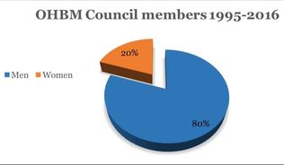
BY THE OHBM DIVERSITY AND GENDER TASK FORCE It’s often been said that the best predictor of future history is past history. Thus, after comments at the OHBM town hall meeting in Geneva regarding the current gender imbalance of Council members (1 female, 14 males), the Council made a decision that something needed to be done to enhance gender equity and geographic diversity. Thus, the OHBM Diversity and Gender Task Force was formed. If it is true that the best predictor of future history is the past, then it was important for the committee to obtain a historical perspective of how OHBM has been doing with respect to women in leadership roles. Perhaps the current 14:1 relationship between males and females is merely a dip in what was otherwise a balance in gender. Thus, we took a close look at the distribution of gender within leadership roles and education at the OHBM annual meetings. Like all good scientists, we will let the data speak for itself (See figures below). Of course, like all good scientists, we also like to say ‘in brief…’ the ratio between males to females in leadership positions, awards, and keynote presentations is about 5 to 1 over the history of the organization. The one area in which there appears to be a transition to greater gender equality is in the Keynote lectures. However, such a transition has not been present in Council positions, which was highlighted at the Geneva Town Hall Meeting. The gender distribution of the general membership is not known because this information has not been requested in the past. Similarly, race and ethnicity of the membership is also not known. However, the gender distribution of poster submissions is approximately 50:50. The goal of the Diversity and Gender Task Force is to help work with Council and the program committee over the next year to recommend changes to promote greater diversity, not only regarding gender, but also regarding ethnicity and geographic distribution. The Diversity and Gender Task Force will be using this blog site as one of our forums to communicate our work. We welcome input from the community to achieve this goal. You can share your ideas and suggestions using the comments field on this blog and/or by sending email directly to the Chair of the Task Force, Tonya White at t.white@erasmusmc.nl. We hope that as a consequence of our work those in the future will look back and see an encouraging historical trend, not only in gender, but also in race, ethnic, and geographic backgrounds of the OHBM leadership. And, most importantly, that diversity will translate into even more impactful and positive advances in our field. As the time is upon us to vote for the new incoming members of Council, it is our desire that you vote for the best candidates, considering each individual, irrespective of gender, race, and geographic location. Please vote, your vote counts!
|
BLOG HOME
Archives
December 2020
|