|
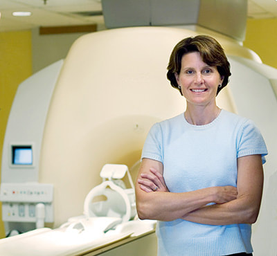
By David Mehler The Local Organising Committee (LOC) at OHBM 2017 achieved a remarkable feat. With public health experts voicing concern about the spread of the Zika virus from South to Central America, it was decided that it was too risky to expose so many young OHBM members to potential infections in Puerto Rico, this year’s original site for OHBM. At this point, the Vancouver LOC stepped forward. They organised an entire, major international neuroimaging conference not in four years, as planned, but in one. Here we speak to Lara Boyd, Professor of translational neuroscience at the University of British Columbia (UBC), chair of the Vancouver LOC, and TEDx sensation. We find out about the challenges of setting up OHBM at such short notice, and about her work mapping out rehabilitative medicine in stroke survivors: David Mehler (DM): It is my great pleasure to introduce Lara Boyd – she’s a professor here at UBC, where she leads the Brain & Behaviour Lab. Lara, perhaps you could give us some insight into chairing the organization of the 2017 OHBM meeting? Lara Boyd (LB): It was an exciting year to chair. In case you missed it, we were supposed to be in Puerto Rico in 2017, and in Vancouver in 2020. Because of circumstances related to world health, we moved it up somewhat suddenly. It only came together through everyone scrambling and working really well together. We were lucky that the venue was available and we were available on short notice. The most fun we had was putting together the local organizing committee symposia, where we could showcase some of the science we do here in British Columbia. That was the best part! DM: Can you tell us what it takes to host OHBM? LB: First, we worked with the organizing committee of the OHBM to find the venue, and select the different speakers for all the symposia; that was a fun process that we really learned from. We worked with the convention centre group just on the physical location; that was less fun but still exciting – particularly in a place that looks like this [looks out the window]. After that, we worked with the student group to make sure we had the venues and the social events planned and they had spaces for Brain Me Out, the Hackathon, those kind of things. Last, we got to put together the symposia. Now, at this point, when we’re all here, we get to just sit back and enjoy and show off a little bit, and that’s been the most fun part. DM: There have been many highlights at the conference so far; for instance, Tal Yarkoni’s input on the statistical implications of fMRI analysis, and then a session on myelin imaging at which you were co-chair. What were your personal highlights of the conference? LB: I loved Tal’s talk --- and I’m not a statistician but he just made that info so accessible. My lab is excited to go home on Monday and try it and see what happens. I also loved the Talairach Lecture, I think Carla Shatz did such a nice job and it’s something I knew nothing about – not my field, not my expertise – so to just sit back and watch how science in one area progressed to something totally unexpected, and she was able to take that knowledge and translate that into something that’s really going to help people with Alzheimer’s and other dementias, that was really exciting. It was just a wonderfully put-together talk. DM: You mentioned translation – your lab is heavily involved in translational neuroscience, particularly in stroke rehabilitation. Your work has contributed to our understanding of how therapies in stroke can work. You’ve more recently shown plasticity even within myelin - very exciting work! It’d be interesting to know what got you into stroke research and what you find particularly interesting about this field. LB: I actually started my professional career as a physical therapist. It didn’t last that long – only about a year. Part of that was because my stroke patients just didn’t get better. I had that sense that I was a car mechanic and didn’t understand how the engine worked. So I went back to school to become a neuroscientist to understand how the brain worked in the hope to translate that information back into therapies for stroke. That’s what led me into the field and it was perhaps good timing, as that was right when the field took off. I believe the first OHBM was in 1995 and that’s when I started my doctorate, so I just grew up with the meeting, and with the field in general. It’s just been a set of really lucky circumstances that has allowed our science and translation to advance so rapidly. I’ve always said that I’m on the consumer end of the neuroimaging spectrum. We take these beautiful approaches that our physicists are designing and we use them to try to really unpack the changes that occur in the human brain. That’s why my lab is called the “Brain and Behavior Lab”. We also try to find out what behavior enables those changes and try to map them. We try to take that information and leverage it into therapies for people with stroke and try to really speed their recovery. We try to enable greater recovery than we’re currently seeing by improving our basic understanding of how the brain changes. DM: At this conference we witnessed that the field of translational neuroscience is rapidly growing. For many young researchers with clinical backgrounds that want to pursue a career in neuroimaging, what would be your top three tips for starting out? LB: First, no question is a dumb question. Don’t be afraid to go up to a senior scientist, or just to someone in an area that you’re unfamiliar with or comfortable with and ask that question. Ask about the field – how did they get into it? What kind of things led them to that? We need to remember that we were all junior scientists and just starting out at one point. I find that everyone is really happy and helpful in sharing their knowledge. Second, make as many connections as possible. You can see the field is incredibly diverse. There are many different imaging platforms. The future I’m seeing is where there is going to be much more multimodal imaging. We can’t be an expert in all of those areas, so we’ll need a lot of good friends. We can start to translate information from different findings in different research studies to understand this marvelously complex thing, the brain. Last, build connections with your peers. These are the colleagues that are going to be reviewing your grants and your papers and in the future these are the people that are going to give you students for your lab as you move along. The more interconnected you can become with your peer group as you rise up through the ranks, the better suited you’ll be when you need that friend who knows a technique when you don’t. You can call upon them and they can really enrich your science. DM: Thanks Lara. Last, can you give us a bit of an outlook for the field of translational neuroimaging for stroke rehabilitation and where you see this field going within OHBM? LB: In stroke rehab right now we’re actually a little bit stuck. Lately, we’ve had a bunch of clinical trials that failed. They failed to show any benefit beyond regular care. In part I think it’s because we treat stroke as if it were a single condition - any of us who have seen stroke patients know that they’re marvelously different. So what we’ve become really interested in, in my group, is understanding biomarkers that can help us sub-categorise people with stroke. We then use those biomarkers to predict what recovery patterns we might see and which treatments are going to be best for which patients. Our stroke recovery biomarkers are all neuroimaging derived. So we can take a human stroke patient, we can use maybe diffusion or myelin water imaging to understand the residual brain structure, understand how that patient may be compensating through different networks in functional patterns, how their cortical excitability is changing with Transcranial Magnetic Stimulation. Then we start to build algorithms and models that take each of those pieces of information and put them together to build a more complete portrait of that patient. We then use that information to predict what may be the best therapy for them. I really think that as we become better consumers of these many different multi-modal types of imaging we can really put them together in a meaningful way. That’s what will move stroke rehabilitation forward, as it will allow us to understand that unique complexity of each patient. It’s sort of what you might think of cancer treatment: cancer treatments are very personalized, highly tailored to each individual. We want to use neuroimaging to do the same thing with our stroke patients. That’s the future I envision and I hope we’re moving rapidly towards it. Thanks Lara, and many thanks to Sarabeth Fox for filming.
0 Comments
Your comment will be posted after it is approved.
Leave a Reply. |
BLOG HOME
Archives
January 2024
|

 RSS Feed