|
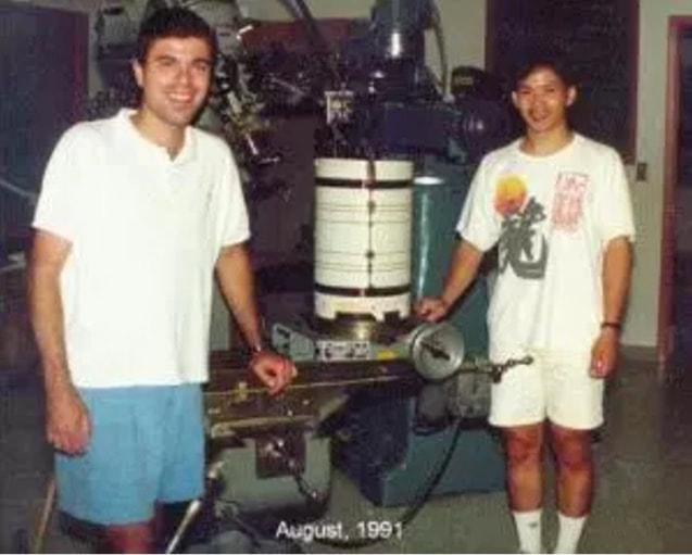
BY GUEST AUTHOR PETER BANDETTINI This post originally appeared on The Brain Blog by Peter Bandettini and Eric Wong. Republished with permission. I’ve been working to advance Functional MRI (fMRI) since its inception. On Sept 14, 1991, two years into graduate school, and a month after seeing preliminary Massachusetts General Hospital results at the SMR meeting in San Francisco, Eric Wong and I performed our first successful fMRI experiment (Bandettini 2012).
Functional MRI, to this day, over a quarter century later, remains as exciting to me as on Day 1 as developments and applications continue at a rapid rate. While human brain imaging methodologies have arisen and grown over the years, and all of them, I’m certain, have interesting stories behind them, I wanted to share why I feel fMRI is unique: 1. fMRI was a surprising, rapid discovery. Elements leading up to the discovery of fMRI were the discovery of BOLD by Ogawa et al (Ogawa 2012), the discovery of the dependence of blood T2 on oxygenation by Thulborn et al (Thulborn 2012), the advent of arterial spin labelling techniques by Williams et al. (Williams, Detre et al. 1992), the technical capability to perform EPI (Cohen and Schmitt 2012), and for the Minnesota group, higher field strengths (Uğurbil 2012). The first fMRI results came from Ken Kwong’s penchant for trying interesting experiments. With Ken’s experiment, the method was discovered rather than incrementally developed. In fact, the pulse sequence and basic parameters used by Ken for BOLD were not anything overly complex or new – simple T2* weighted gradient-echo EPI at 1.5 Tesla. There was minimal time series processing involved then – in stark contrast to processing methods today. Interestingly, aside from the explosion in the sophistication of time series processing, the details of Ken’s first experiment have not qualitatively changed in terms of general practice over the years. He was just the first to realize that such a straightforward thing could be done! This discovery surprised and excited the MRI community. To provide an analogy, it was as if we realized that if one sets the exposure settings of a standard camera just right, rather than just getting a photograph, you can get a picture of, say, subatomic particles. While the MRI scanner vendors adopted a wait-and-see approach before putting any resources into developing fMRI, the clinical and basic neuroscientists were highly motivated to start scanning. 2. fMRI was a revolutionary advance in functional imaging capability. Functional MRI was, and still is, the only non-invasive, whole-brain method that has enough sensitivity to see human brain activity with about 2mm detail as it is happening in real time. This made for good science fiction before 1991. No one imagined it would become reality so quickly. 3. fMRI is deeply multidisciplinary. Functional MRI brought disparate disciplines together in a way that was unprecedented. Suddenly, cognitive neuroscientists were having intense conversations with MR physicists. Computer programmers were talking with clinicians. The best fMRI research today has a signature of advancing methodology and insight into brain function – requiring close collaborations between physicists, statisticians, programmers, and neuroscientists. 4. fMRI is riding on the back of the clinical MR industry. A huge factor that many people overlook is that fMRI was able to launch and propagate so rapidly because it leveraged the massive clinical MRI industry. In the early 90’s there were at least 20,000 clinical MRI scanners worldwide. By 1998, most MRI scanners were equipped with EPI – for other more clinically relevant purposes such as following a bolus of gadolinium for perfusion imaging or visualizing the heart beating. Even though fMRI had minimal clinical impact, almost every MRI scanner in every hospital in the world was a potential brain function imaging machine. There was no need for a manufacturer to make fMRI machines. They already existed! These scanners were priced at over $1M each and were paid for and supported by hospital revenue – not neuroscience research grants. Today, that’s changing somewhat as the fMRI market grows and research grant revenue towards fMRI increases, but the reality is that fMRI depends on the clinical MRI market to survive. fMRI has tremendously benefited from essentially riding on the back of the clinical MRI industry. This relationship has clear drawbacks too. Many interesting pulse sequences and custom fMRI setups are not being disseminated worldwide because the scanner vendors do not yet see a large enough market of fMRI to necessitate adding more development resources. Until fMRI becomes a thriving clinical technique (hopefully soon), it will be at the mercy of the clinical focus of the MRI scanner vendors – namely Siemens, General Electric, and Philips. 5. The degrees of freedom in fMRI acquisition is vast and unexplored. We can do so much more than collect a simple time series of T2* weighted echo planar images. The ability to derive physiologic and neuronal information from MRI is still being explored as there are so many “knobs” you can adjust on the acquisition side to highlight gray matter, white matter, CSF, flowing blood, perfusion, iron deposits, vascular territories, trauma, leaks in the blood brain barrier, hemorrhage, deoxygenated blood, metabolism, pulsation, macromolecules, temperature, water diffusion, diffusion anisotropy, and much more. Additionally, the information that may be useful to fMRI is also still relatively untapped. Along with mapping the magnitude of the hemodynamic response as is most commonly done, we can derive information about latency, fluctuations, oxidative metabolic rate changes, blood vessel sizes, oxygenation, and more. 6. Processing methods are exploding in variety and sophistication. Functional MRI processing methods continue to surprise – as it seems that the field continues to find new and better ways to extract, compare, and display new hemodynamic and neuronal information in groups and individuals. With the emergence of massive shared data sets, ever more subtle information about individual differences and similarities is being plumbed with the help of modern machine learning approaches. 7. Functional MRI just works. Functional MRI just works – almost every time! It’s a stunningly robust technique. The functional effect size to noise ratio (from 6/1 to 1/1) is still perhaps too small and subject-wise variability a bit to large (with current post processing techniques) for robust clinical use (at least 10/1 is considered essential) but is large enough to see a significant effect within a few minutes of averaging. If it took 6 hours of averaging to see something, ambitious people would still do it but it would be much more difficult and the field would be much more anemic at this point. 8. fMRI requires two highly serendipitous properties. Another key to fMRI that is commonly taken for granted: It requires two very subtle yet all-important properties to be possible at all. The first is that hemoglobin has to change its magnetic susceptibility in a non-trivial manner between being oxygenated and deoxygenated. This is an extremely rare property of a biologic tissue. If our blood were copper based – as with mollusks – rather than iron based, this would not happen. We would not have BOLD contrast as copper based blood does not change susceptibility with oxygenation. The second all-important property is that, with activation, a localized flow increase in the active region creates a highly focal overabundance of oxygenation. Why didn’t nature just require that the oxygenation stay the same in the active regions? We are still trying to figure that out, but the fact that it does – every single time with every person – similarly across species – in the same precise way in a stunningly consistent manner is highly fortunate. Perhaps our brains could have evolved a system where localized activation-induced changes in flow increased to simply match the increased metabolic needs rather than apparently overshoot them. If this happened, there would be no BOLD changes. We are lucky! 9. fMRI fills a unique temporal and spatial niche. The information that fMRI provides fills a large and interesting temporal and spatial niche in understanding brain organization. Our brains are highly modular, and fortunately, the larger modules (motor cortex, visual cortex, etc..) are easily large enough to be discerned with fMRI. If our largest brain modules happened to be no larger than ocular dominance columns, fMRI would have never taken off, and if it did, interpretation of the results would have been a challenge at best. We’ll likely gain enough sensitivity and resolution soon to routinely probe the columnar and layer level organization of the brain soon – which brings us to the next unique property… 10. The highest fMRI spatial resolution matches the intrinsic precision of hemodynamic control. It appears that the highest resolution achievable to fMRI (limited by scanning technology) – that of cortical columns or layers – matches the intrinsic precision of hemodynamic control. In other words, the smallest homogeneously activated region that causes a focal change in blood flow is on the order of columns or layers (<1mm). This perhaps suggests that this is the smallest scale in which groups of neurons are activated together. This last point is potentially controversial as it may suggest that looking any finer than this scale at neuronal activity may not necessarily lend insight into modular brain organization. Either way, again it’s fortuitous that fMRI resolution limits match the hemodynamic control limits – at least in humans. The OHBM Blog Team thanks Peter Bandettini for this guest post. Interested in submitting a guest post for consideration? Email your post to the OHBM blog team at [email protected].
0 Comments
Your comment will be posted after it is approved.
Leave a Reply. |
BLOG HOME
Archives
January 2024
|
 RSS Feed