In this week’s podcast, Dr Catie Chang walks us through her thought process regarding pulling information out of the fMRI time series. After discussing some of the ongoing issues in fMRI, such as whether or not to use global signal regression to remove noise, she leads us into a commonly overlooked effect in fMRI—that of changes in arousal and vigilance. In particular, this has measurable effects on the resting state fMRI signal. She discusses the perspective that one person’s artifact may be another’s useful signal, depending on the goal of the study.
Guest:
Catie Chang, Ph.D. received her B.S. in Electrical Engineering and Computer Science from MIT, and received her M.S. and Ph.D. in Electrical Engineering from Stanford University. While in graduate school, she opened up the field of fMRI by publishing a seminal paper using time-frequency analysis of resting state fMRI, showing that it was quite dynamic. Since then, she has been exploring the effect of basic physiological processes, such as cardiac function and respiration on the fMRI signal, and has recently been uncovering unique information regarding the influence that changes in vigilance have on the time series signal. --- The Neurosalience production team consists of Rachael Stickland, Kevin Sitek, Katie Moran and Anastasia Brovkin
0 Comments
By Peter Bandettini & the OHBM Neurosalience production team
In this week's episode, Peter talks to directly to MRI scanner vendors. Together, they try to reconcile the importance of fMRI in research contexts with the market pressures of developing clinical applications. As fMRI has virtually no clinical market, does it really influence vendor decisions on pulse sequences and hardware? Could more be done aside from making fMRI more clinically relevant? In this discussion, you’ll hear some fascinating history into the early days of echo planar imaging and high speed imaging, as well as insight into the processes by which products are prioritized. You’ll also find out a possible future of how fMRI may begin to become more clinically useful. By Charlotte Rae, on behalf of the SEA-SIG The Sustainability and Environment Action (SEA) SIG has formed three new Working Groups, to tackle the environmental impact of the annual meeting, assess environmental implications of neuroimaging research activities, and educate our community on these. What are the new Working Groups? In December 2020, we held two open meetings to talk about the priority actions for our new SIG with the OHBM community. We had colleagues attend from across the world, who shared fantastic ideas on how we should make OHBM activities more sustainable. From these meetings, there was a pretty clear consensus that we needed to tackle three areas: the Annual Meeting, neuroimaging research pipelines, and education. So, we have set up three new Working Groups that will focus on these particular domains. The Annual Meeting Working Group will assess the environmental impact of the Annual Meeting, investigate sustainable conference models, and make recommendations to the Council for how to create a more sustainable Annual Meeting beyond COVID-19.
By Peter Bandettini and the OHBM Neurosalience production team
In this week's podcast, Peter gets a birds-eye view of modeling of messy biologic systems, namely the brain, from Professor Danielle Bassett. They talk about the challenges of measurement accuracy and what scale might be most informative to modeling - and how to make do with what we have. On the clinical side, Danielle discusses network control theory for modulating networks for therapy and limitations in technology for modulation. They consider the limits of network modeling and the search for the equivalent of an idea as powerful as “natural selection” for the brain. In the second part of the podcast Peter and Danielle discuss bias in science and what Danielle is doing to help increase transparency to combat bias.
Danielle Bassett PhD, is currently the J. Peter Skirkanich Professor at the University of Pennsylvania with a primary appointment in the Department of Bioengineering and a Secondary appointment in the Departments of Physics and Astronomy, Electrical and Systems Engineering, Neurology, and Psychiatry. Dr. Bassett received her B.S. in 2004 in Physics from Penn State University. She received a Ph.D. in physics in 2009 from the University of Cambridge, UK as a Churchill Scholar, and an NIH Health Sciences Scholar. Following a postdoctoral position at UC Santa Barbara, she was a Junior Research Fellow at the Sage Center for the Study of the Mind. In 2013, she joined the University of Pennsylvania as an assistant professor, and in 2019, was promoted to full professor. She is also founding director of the Penn Network Visualization Program, a combined undergraduate art internship and K-12 outreach program bridging network science and the visual arts. Her primary work is towards developing network models towards deriving principles of brain function. The Neurosalience production team consists of Rachael Stickland, Kevin Sitek, Katie Moran and Anastasia Brovkin
By the Neurosalience production team: Rachael Stickland, Kevin Sitek, Katie Moran and Anastasia Brovkin
 Our Neurosalience logo, designed by Roselyne Chauvin Our Neurosalience logo, designed by Roselyne Chauvin
OHBM has a new podcast: Neurosalience! You can listen to it in your car, while out walking, or just in the ever-present home office. Through Neurosalience, you’ll learn about recent advances and current controversies in brain mapping. The host for the podcast, Peter Bandettini, has lined up a stellar cast of interviewees ranging from brain scientists to hardware vendors and health professionals. This includes finding out about publication biases affecting gender and racial minority groups with Dani Bassett, network neuroimaging in neurological populations from Michael Fox, circuit based neuromodulation from Catie Chang and much more. Get all of this insight through your favourite podcast apps, including Spotify, apple podcasts, anchor and Google Podcasts.
We launch with a brief introduction to the podcast, a fireside chat between Peter and Rachael Stickland (one of the OHBM Communication Committee producers for the show). Then the first full episode explores Aperture, the new open-access publishing platform powered by the OHBM. Through discussions with founding members and the Editor in Chief, you’ll learn how Aperture came about and what it hopes to achieve. 2020 was such an interesting year; it was certainly not the one I was waiting for. Due to several issues related to the pandemic, I unofficially took a leave from thesis work and had a chance to meet a lot of people virtually, collaborate, learn and grow. Although so many of us were stuck at home, open-science-driven events like NeuroMatch and BrainHack created opportunities to connect with colleagues and peers. This turned out to be hugely impactful for myself and other people like me—in other words students/early career researchers based in countries with limited resources. And to think, it all started with a tweet…. 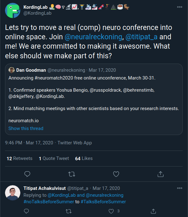 Figure 1. Neuromatch 1.0 call for people (image credit: https://twitter.com/KordingLab/status/1239986383550365696).
 Professor Helen Mayberg Professor Helen Mayberg By Rachael Stickland & Nils Muhlert
Professor Helen Mayberg is a pioneer of neuroimaging and neurostimulation for depression. As a behavioral Neurologist she has helped to identify the brain circuits implicated in mood disorders, and then developed and refined effective treatments based on deep brain stimulation. She is a member of the National Academy of Medicine, The American Academy of Arts and Sciences and the National Academy of Inventors. As a founder member of OHBM we found out about her work, her experiences of seeing impact statements become reality and about holding on to the ‘OHBM train’. Ilona Lipp (Lead editor):
By Valentina Borghesani, Elvisha Dhamala, Niall Duncan, Marie-Eve Hoeppli, and Michele Veldsman, on behalf of the SEA-SIG
This month, OHBM announced the formation of a new Special Interest Group that will tackle sustainability and environmental issues around brain imaging. Here, we talk with the Sustainability & Environment Action (SEA) SIG Chair Charlotte Rae to hear more about what the new SIG will seek to achieve. By: Rosanna Olsen, Amanpreet Badhwar, Valentina Borghesani, Lee Jollans, Hajer Nakua, Laura Marzetti, Nils Muhlert, Pradeep Reddy Raamana, Tilak Ratnanather, and Lucina Uddin on behalf of the OHBM Diversity & Inclusivity Committee
In June 2020, OHBM made a statement condemning the murders of George Floyd, Breonna Taylor, and Ahmaud Arbery as well as ongoing actions of police brutality against Black Americans and underrepresented minorities around the world. During the conversations surrounding these events, there was a public recognition of the lack of support for Black and minority communities. We realized that at OHBM we have not done enough to support underrepresented minorities in science, and that we need to take concrete actions to make our organization a welcome and safe environment that educates and supports each and every member of our group. by Claude Bajada
The GDPR is a new(ish) legislation by the European Union that regulates the processing of personal data when the person processing or controlling the data is in the EU, even if the actual processing occurs outside of the EU. Further, the GDPR also sometimes regulates the processing of personal data of people who are in the EU, even if the persons doing the processing are outside of the EU. How does this affect neuroimaging? We sit down with neuroimaging expert and Open Brain Consent co-author Dr Cyril Pernet (CP) and Technology law expert Dr Mireille Caruana (MC) to discuss the implications of this law on our work. The article flip-flops between the term “participants” and “data subjects” since ““data subject” is the term used in the GDPR but for the purposes of this article you can think of them as equivalent terms. What follows is a summary of our conversation, edited for conciseness and clarity. Who are our experts? Now is the time to submit your nominations for 2021 OHBM Awards. To inspire you, we are highlighting some of the outstanding winners from this year’s meeting.
This year’s annual meeting was unique in many ways. Uncertainty about whether the meeting would happen was followed by a remarkably fast reorganization in order to hold the meeting online with a complex time schedule. One event that was not missing in the program was the traditional award ceremony that recognized the work of individuals who have changed the scientific landscape of human brain mapping. Inspired by their nomination letters, we honor OHBM 2020 award winners and their achievements: Written by: Claude Bajada, Fakhereh Movahedian Attar, Ilona Lipp
Expert reviewers: Adina Wagner, Cyril Pernet Newbie editors: Yana Dimech, Renzo Torrecuso This post is about good neuroimaging practices. ‘Practices’ relates to all aspects of conducting research. By ‘good’, we mean beneficial to the field and neuroimaging community - but you’ll see that most of these practices also benefit the individual researcher. Here, we collected a number of tools, tips and tricks to do neuroimaging in the ‘best’ way possible. We aim to provide an overview and answer some questions you may have asked yourself about reproducibility and good neuroimaging practices. As usual, we refer to OHBM On-Demand videos from the educational sessions of previous annual meetings. OHBM has both a special interest group (SIG) for Open Science as well as a Best Practices Committee, where leading brain mappers promote and help implement Open Science and good practices in data analysis and sharing. Both the Open Science SIG and the Best Practices Committee regularly create invaluable resources, such as the annual Hackathon workshops, and the COBIDAS Best Practices in MRI and M/EEG data analysis papers. Guest post by Hiromasa Takemura
International diversity is essential for organizations like OHBM. Through my experiences attending recent OHBM Annual Meetings, I have found myself asking why so few researchers from Japan have visible roles. To find out whether this was indeed the case, and possibly why, I worked with the OHBM Executive Staff, Diversity & Inclusivity Committee, and Communications Committee to analyse membership and attendance data from the annual meetings. By collecting and analysing this demographic data we can gain insight into why some countries (in this case Japan given my background, but the findings may extend to others) may be underrepresented at OHBM. Japan is the 11th most populous country in the world, with an estimated population of 126 million (m) people in 2020. For comparison, Mexico has the most similar population with 128m people and Germany, Europe’s most populous country, has 83m. Japan has, over the years, substantially contributed to the OHBM community: for instance, the 2002 Annual Meeting was held at Sendai, Japan and Dr. Kang Cheng, a pioneer of high-resolution fMRI studies at a founding lab for RIKEN's Brain Science Institute, is heavily involved in organization of OHBM meetings. To get a picture of recent involvement of researchers from Japan, we examined data summarizing attendance and presentations at the OHBM Annual Meeting between 2017-2019 (Table 1). We defined Japanese members as those affiliated with Japanese institutions. Using this definition we found that Japanese members comprised 3.6%, 5.4% and 3.9% of all attendees for 2017, 2018, and 2019 respectively, with the fluctuation reflecting the location of the annual meeting (OHBM 2018 was held in Singapore). We found a lower proportion of abstracts submitted by Japanese members: 2.6%, 3.6%, and 3.6% of the total number of abstracts for each of these years. By Elizabeth DuPre
The OHBM 2020 Annual Meeting was a year of many firsts. The move to an all-online event reflected the severity of the COVID-19 pandemic, with work, travel and schooling routines already in disarray for researchers across the globe. As many of us had been out-of-office or away from our university campuses for months before the Annual Meeting, the chance to connect with the broader human brain mapping community became especially important. Traditionally, the Annual Meeting offers a chance to interact formally and informally with other researchers to make both scientific as well as interpersonal connections. Replicating these spontaneous conversations was perhaps the biggest challenge for this year’s meeting. First, there were the issues of timing. With OHBM members participating from their home countries, one member’s afternoon in North America would be the middle of the night for another member in Asia. The meeting was therefore set on a rotating schedule, with day-blocks favoring Asia-Pacific, European and African, or North and South American working hours. Once the timing was set, the second hurdle was developing a virtual space for interactions. Large online platforms—like those necessary to run a conference for thousands of members—often lean towards structured, lecture-style environments rather than organic interactions and impromptu discussions. From the available infrastructure options, OHBM Council decided in April to adopt the 6connex platform. Council’s intention was to allow time for all presenters, committees, and special interest groups (SIG) members to adapt their content; however, the time pressures of the COVID-19 pandemic meant that many were still unclear how this new platform would work in practice in June. Expectations were thus high for the 6connex platform—possibly higher than could be reasonably met. The platform did well in delivering pre-recorded content, such as the excellent selection of keynotes lectures, symposia and oral sessions, but the space for spontaneous interaction was woefully lacking. As one example, many members noted the challenges of using the chat feature, such as when 1000+ attendees simultaneously participated in a single-threaded chat room. This lack of functionality created particular frustration in poster presentations and interactions, where presenters and attendees were unclear how to contact one another or how to provide on-the-spot poster walk-throughs. By Tzipi Horowitz & Nils Muhlert
Institutions throughout the world have had to adapt to the Covid-19 pandemic. Many scanning centres shut their doors during lockdown, and have had to reopen gradually, and carefully. We surveyed several labs from around the world - to find out the challenges they’ve experienced and, in a few cases, the opportunities afforded. UK - Matt Wall (Head of MRI applications, Invicro, Hammersmith Hospital, Imperial College London) Challenges: Everything shut down rapidly at the start of lockdown. In March, two big commercial scanning projects had to stop immediately. One had been running for some time, the other had just started. We had a lot of clinical people working with us - some very good medics. They spent a lot of time developing risk assessments and procedures. So we ended up restarting in late June. I tweeted about it at the time, and was contacted by people in other universities, asking how we managed it - so we shared our findings from the risk assessment process. 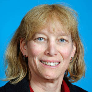 Tonya White Tonya White By Nabin Koirala
In advance of our scheduled launch of the upcoming Journal “Aperture” from the Organization of Human Brain Mapping (OHBM), we wanted to get up close with the first Editor-in-Chief of the Journal - Tonya White. Tonya is currently an Associate professor in the Department of Radiology and Nuclear Medicine and Department of Child and Adolescent Psychiatry in the Erasmus Medical center in Rotterdam, Netherlands. We discussed her personal journey in Science and her vision for the Journal. Nabin Koirala (NK): Thank you so much for making time for this interview. To start, could you please introduce yourself to general readers who may not be scientists? Tonya White (TW): That's always an interesting question because I have a number of different hats. I could say that I'm a developmental neuroscientist, a child and adolescent psychiatrist, a pediatrician or an electrical engineer. But what I've been mostly doing currently is what's called pediatric population neuroscience, which is actually the intersection between epidemiology and developmental neuroscience. The neuroimaging program I came to set up in Rotterdam is a large population-based study of child development. We’re currently collecting and evaluating more than 8000 MRI scans from children at three time points. Through the so called “Generation R Study” nearly 10,000 mothers who were pregnant between 2002 and 2006 were approached to participate in the study and the imaging is nested into a multifaceted epidemiologic study looking at many different aspects of child development. 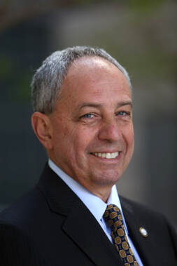 John Mazziotta is Professor of neurology, CEO of UCLA Health, and vice chancellor of UCLA health sciences. He was also a founding member of the OHBM. He co-authored the first book on whole-body, cross-sectional anatomy using CT. He’s been involved in the first PET studies in normal subjects and with patients with epilepsy and Huntington’s disease. He was the principal investigator of the ICBM brain atlas, a key tool for brain normalisation. We interviewed him as part of our OHBM Oral History series, to find out about the early days of PET, (f)MRI and the inception of OHBM.
Nils Muhlert (NM): Thank you very much, Professor Mazziotta, for joining us today. I'd like to start by asking you about your background: Why and how did you become interested in neuroimaging? John Mazziotta (JM): Well, I wanted to be an architect. That didn't work out because I spent a lot of time in Manhattan with architects when I was an undergraduate, and they didn't seem very happy. I like science and went into a lab where I was doing early molecular biology and that was interesting but very isolating. I thought, “Well, I'll go to medical school.” I hated medical school, memorizing bones and things of this sort. Ultimately, I met a neuroscientist in the medical school. The school also had a very active biophysics department and were building the first CT scanner that could image outside of the head. This is now mid-1970s. I got involved in that project and we physically built that machine, soldering wires. We had a functional scanner that worked anywhere in the body. I decided I would become a neurologist, moved to Los Angeles and UCLA and immediately met the group that had moved from Washington University in St. Louis. They had been involved in the development of PET and all worlds connected, so I got involved in research with PET and then MRI. Authors: Katie Williams, Ilona Lipp, Mark Mikkelsen Infographic: Roselyne Chauvin Expert editors: James Kolasinski, Paul Mullins Newbie editors: Curtiss Chapman, Yana Dimech Introduction
The noninvasive imaging tools that we Human Brain Mappers apply are most often being used to research brain structure and function. Neurotransmitter systems are something that we are aware of and use to take into account when coming up with hypotheses or interpreting our findings, but rarely make the direct subject of our investigation. Most of us have probably heard of GABA (gamma-aminobutyric acid) as the principal inhibitory neurotransmitter that is used by many interneurons. That we can also measure GABA in vivo with MR spectroscopy (MRS) is maybe less widely known. While this biomedical imaging tool opens many doors for neuroscience, measurement of GABA using MRS is not broadly used yet, possibly because special sequences and analysis methods are needed. At the OHBM Annual Meeting in 2019, for the first time, an educational session on GABA MRS was held. This post summarizes what was taught about the most important things you need to know if you’re considering GABA MRS for your research. by Nikola Stikov As we are getting ready to announce the 2020 OHBM Replication Award winner, here is a brief flashback to 2019 and our interview with Richard Dinga from the Department of Psychiatry at the Amsterdam University Medical Centers in the Netherlands. Richard led the effort to replicate a study published in Nature Medicine in 2017 about the relationship between resting state connectivity and the neurophysiological subtypes of depression.
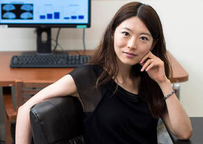 Biyu He Biyu He In the lead up to the OHBM Annual Meeting, I had the pleasure of speaking to one of the keynote speakers, Dr. Biyu He, an Assistant Professor at New York University. Dr. He has made many valuable contributions to the field of neuroscience, combining diverse imaging methods and analytical techniques to tackle big questions relating to perceptual processing, spontaneous activity and consciousness in the human brain.
Rachael Stickland (RS): Thanks again for joining me. It's nice to - virtually - meet you. Biyu He (BH): Pleasure to meet you as well. OHBM 2020 Diversity Round Table: Intersection between Neuroscience and the LGBTQ+ community6/18/2020 Lee Jollans and the OHBM Diversity and Inclusivity Committee.
Edited by AmanPreet Badhwar At the 2020 virtual meeting, OHBM will, for the second time, host a Diversity Round Table. This year the round table will feature discussions on the intersection between Neuroscience and the Lesbian, Gay, Bisexual, Transgender, and Queer (LGBTQ+) community. The four speakers will outline the specific challenges LGBTQ+ individuals face working in STEM (Jon Freeman), insights into the possible developmental bases of sexuality and gender (Doug VanderLaan), the current body of research into transgender identity (and its limitations), and the challenges and considerations that are crucial for carrying out good sex and gender research (Grace Huckins and Jonathan Vanhoecke). 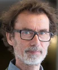 Tomas Paus Tomas Paus In preparation for OHBM 2020, we talked to Dr Tomas Paus, who will be giving a keynote lecture on Friday, June 26th. Dr. Paus is Director of the Population Neuroscience & Developmental Neuroimaging Program at the Holland Bloorview Kids Rehabilitation Hospital, and Professor of Psychology and Psychiatry at the University of Toronto.
Roselyne Chauvin (RC): Thank you for taking the time to chat with us. In your talk you will be speaking about “population neuroscience and the growing brain.” There are a few ongoing longitudinal big data initiatives, such as ABCD or generation R. Those projects are now starting to think about the current pandemic situation. On one side, the situation is affecting everyone without discrimination; on the other, government responses create different experiences (from full to partial lockdown, to no restrictions), and of course, individuals show different stress responses. How do you think this might affect longitudinal datasets? And what are the questions that will need to be investigated out of this situation with regard to psychiatry and genetics? By Nils Muhlert  Michael Fox Michael Fox Professor Michael Fox is a neurologist at Harvard Medical School and director of the Lab for Brain Network Imaging and Modulation. His research into brain network imaging to define targets for brain stimulation holds considerable promise for new and improved treatments for a wide range of neurological and neuropsychiatric conditions. Here we found out how his academic career started through a chance meeting with Mark Raichle, about his plans for clinical translation of network neuroimaging, and his advice for early career researchers:
By the OHBM Diversity and Inclusivity Committee (and endorsed by OHBM Council)
We share the deep sadness, outrage, and frustration that many around the world have felt in reaction to the murders of George Floyd, Breonna Taylor, and Ahmaud Arbery, and too many other innocent Black people over the years. As an international organization that strives to represent a diverse and vibrant global community of researchers studying the human brain, OHBM itself has struggled over its 25 years to incorporate initiatives and policies that reflect our values of inclusivity, tolerance, and respect. The events of these past weeks are a grim reminder that words alone are not enough to combat the systemic racism that plagues societies across the world, and we recognize that we have not done enough to support Black, Indigenous, and People of Color. |
BLOG HOME
Archives
January 2024
|
 RSS Feed