|
By the OHBM Communication Committee
By now you've heard that the OHBM Annual Meeting will be virtual! The 26th Annual Meeting of the Organization for Human Brain Mapping is happening from June 23 - July 3, Saturday and Sunday excluded, and will take place entirely online. This is new for many of us so we’ve put together a short Q&A. Here we address a number of questions you may have, and provide a taste of what you can expect from this unique OHBM Annual Meeting experience.
0 Comments
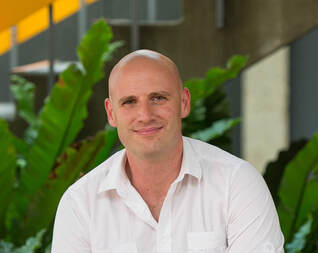 Alex Fornito Alex Fornito Of course we all know that the brain functions as a network, but it is not straightforward to model it as such. One person who works very hard for us to be able to do so is Alex Fornito. He is a professor at Monash University and one of the leading forces in MRI-based network neuroscience. As he is also one of this year’s virtual meeting’s keynote speakers, I had the pleasure to invite Alex to a virtual meeting to ask about his scientific life.
Ilona Lipp (IL): Thanks for joining me during these crazy times. Apart from OHBM going virtual, what else has changed in your scientific life in the last few weeks? by Athina Tzovara, Julia Kam, Valentina Borghesani, AmanPreet Badhwar ‘If you never did you should. These things are fun and fun is good’ - Dr. Seuss
Live Review for Kids The OHBM Diversity and Inclusivity Committee is exploring an exciting and new direction this year: we will be engaging kids in the scientific review process! We asked five prominent scientists in the field of brain mapping and neuroscience to write a short article explaining their research to kids. The articles are written for the Frontiers for Young Minds (https://kids.frontiersin.org/, a journal dedicated to young readers of 8-15 years old. Once written, the articles are assigned to five young reviewers, who will work together with a neuroscientist mentor to critique the articles and prepare questions for the scientists. 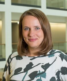 Claudia Buss Claudia Buss by Ekaterina Dobryakova
In preparation for this year’s Annual Meeting, we spoke to one of the keynote speakers, Dr. Claudia Buss. Claudia is an Associate Professor at the University of California, Irvine and a Professor of Medical Psychology at the Charité University Medicine in Berlin. In a virtual meeting, I sat down with Dr. Buss to discuss her captivating research in the field of developmental programming and newborn infant neuroimaging. Ekatarina Dobryakova (ED): Dr. Buss, thank you for dedicating your time for this interview. Before we get into more specific questions, I was wondering whether you mind sharing a bit about how you came to do the work that you're doing, and what got you to follow this passion in research. Authors: Claude Bajada, Nils Muhlert, Ilona Lipp
Infographic: Roselyne Chauvin Expert editors: Alfred Anwander, Jurgen Gatt Newbie editor: Caroline Jantzen Introduction Neuroanatomy is one of the most exciting topics in neuroscience! Some readers may disagree, but for now, humor us and read along. With the help of this On-Demand post, we will convince you not only that anatomy is a useful endeavour but that it is one where much beauty is found. Our journey starts with the fundamental notion that the structure and the function of objects are tightly coupled; sometimes in ways that are not obvious. Understanding the complexity of the brain’s structure, hopefully, allows researchers to build more accurate models of brain function. 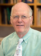 Professor David van Essen Professor David van Essen David van Essen, Alumni Professor of Neuroscience at Washington University St Louis School of Medicine, has been a pivotal figure in non-human and human neuroimaging. David is the principal investigator on the Human Connectome Project, and has made substantial contributions to brain parcellation methods, functional neuroimaging, and data sharing initiatives. Here, we find out about his early work in cortical cartography and his early experiences with OHBM.
 Aperture, OHBM’s exciting new open-access publishing platform, is on track for a June launch, just in time for the 2020 Annual Meeting. The Aperture Oversight Committee, consisting of Jean-Baptiste Poline, Peter Bandettini, Michael Breakspear, Nikola Stikov, David Kennedy, and Jessica Turner, has been hard at work finalizing a plan of operations that will help guide all of Aperture’s submission, editorial, and review processes.
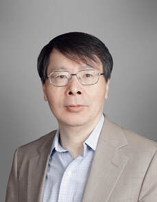 Jia-Hong Gao Jia-Hong Gao By Lisa Nickerson
The current Chair of the OHBM is Jia-Hong Gao, who brings a fresh perspective being the first Chair elected from Asia. Jia-Hong received his Ph.D. from Yale, followed by post-doctoral work at MIT and faculty positions at San Antonio and Chicago. Since 2013, Jia-Hong has been in Beijing as the Director of the Beijing City Key Lab for Medical Physics and Engineering and a Principal Investigator at the McGovern Institute for Brain Research at Peking University. We interviewed him about his experiences as Chair of OHBM, what excites him most about neuroimaging, and the rapid expansion of neuroimaging research in China. By Nils Muhlert “The times, they are a-changing.” Dylan’s lyrics from the early 1960s reflected that era’s mass societal upheavals. But now, with increasing realization of the impact of climate change, with a once-in-a-generation response to the dangers of a pandemic, those words ring truer than ever. Life is definitely moving apace. But our work in trying to understand the structure and function of the human brain continues. These efforts to improve scientific methods, make more accurate insights and accelerate the communication of that work remains.  Balancing the budget requires trade-offsBy Lilianne R. Mujica-Parodi (OHBM Treasurer 2019-20) At OHBM 2019, Council decided that it would be beneficial to the membership to provide a window into the decision-making process regarding finances. If you’ve ever wondered why our support for Special Interest Groups (the SIGs) changes from year to year, or how we decide on the location, venue, and registration costs for a meeting—we hope to demystify some of the many thought processes that go into how Council makes its financial decisions and prioritizes requests for funding.
Responsible financial stewardship of OHBM has always been a priority of the Society. This includes the maintenance of adequate financial reserves that are needed for a society to function. For OHBM, this requires that our financial reserves are equal to at least 50% of the average annual costs, averaged over the previous three years. This is consistent with professional investment advice, and how many societies run their finances. By Valentina Borghesani & AmanPreet Badhwar
And here we are, January 2020! A new decade is starting, with end-of-the-year reflections giving way to wishes and resolutions for the future. As Chair and Secretary of the BrainArt Special Interest Group (SIG), the youngest SIG within OHBM, we will here outline our inner structure to introduce you to our members, briefly cover our past endeavors, and finally sketch our plans for the coming year. Buckle up! The coming of age of OHBM Brain & Art initiatives For us, 2019 has been the year of the transition from a rather informal group of aficionados, supported and sustained by the Neurobureau, to a structured team within our Society. Last year's meeting in Rome thus signed not only the 25th anniversary of our community get together, but also our graduation, so to speak [you can read more about our transition here]. BY NILS MUHLERT (Lead editor)
The OHBM blog is now entering its fifth year. In that time it’s moved from primarily an interview-based format to embrace a diverse range of educational and entertaining posts. In 2019 this culminated in the introduction of the popular ‘How to’ series. This leveraged the rich content of previous OHBM lectures to teach novices, intermediates and experts about setting up and analysing resting-state fMRI and diffusion MRI studies, and using machine learning in neuroimaging. Spearheaded by Ilona Lipp, our Chair Elect for the Communications Committee, these painstakingly crafted posts offer one of the best freely available resources for those wanting to get to grips with these techniques. We also managed to shine the spotlight on neuroimaging efforts in China and Iran, and plan to continue highlighting brain mapping across the globe throughout the ‘roaring 20s’. Finally, my personal highlight from 2019 was to interview Bruce Rosen. He offered not only deep insight and historical perspectives of fMRI but did so with good humour. Looking forward to more next year. 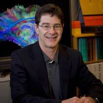 Professor Bruce Rosen Professor Bruce Rosen By Nils Muhlert
Bruce Rosen is a physicist and radiologist who, for the past 30 years, has been instrumental in the introduction and development of functional MRI. Bruce currently serves as the director of the Athinoula A. Martinos Center for Biomedical Imaging at the Massachusetts General Hospital. Here, we found out about the exciting early stages of putting a team together to discover and develop the principles of fMRI, and helping to found OHBM in the process.  Jeanette Mumford Jeanette Mumford By Ilona Lipp
Every year, the OHBM gives out an Education in NeuroImaging Award, acknowledging significant contributions to education and training in our field. This year’s award went to Jeanette Mumford. Jeanette is a well-known fMRI stats guru who spreads her knowledge not only through her published papers but also through YouTube and Facebook, and in the handbook for fMRI analysis. Many of you may also have tried her fMRI power analysis tools. I had the pleasure of meeting Jeanette in Rome and interrogating her about how she became such an enthusiastic educator and her views on neuroimaging research. By Bin Lu and Niall Duncan
Recent years have seen a number of important themes come to the attention of the global neuroimaging community. The robustness of findings reported in the literature have been questioned as people begin to focus more on reproducibility and other statistical issues. At the same time, more attention is being paid to the variability between individuals, not least as efforts to develop diagnostic tools for different brain diseases advance. Databases of imaging data from very large samples have come to the fore as one way of tackling these issues and have already led to some striking results. Israel is a small country, approximately 400 km long north to south and 25 km width at its narrowest point. Despite its small size, Israel is home to six large universities and this year hosted the 1st Human Brain Mapping conference. This inaugural conference aimed to bring together neuroimaging researchers from each of these universities, to share ideas and methods. The conference unites those working on a number of different modalities - as was shown by the diversity in over 70 talks and posters, with research using MRI, fNIRS, MEG, EEG and brain stimulation, studying populations across the lifespan.
By Johannes Algermissen, James Bartlett, Remi Gau, Stephan Heunis, Eduard Klapwijk, Matan Mazor, Mariella Paul, Antonio Schettino, David Mehler The neuroimaging field has recently seen a substantial surge in new initiatives that aim to make research practices more robust and transparent. At our annual OHBM meetings you will have likely come across the Open Science room. While many aspects fall under the umbrella term Open Science, for this post we focus on research practices that aim to make science more replicable and reproducible. These include non peer-reviewed study preregistration, peer-reviewed registered reports that reward researchers’ study plan with in-principle acceptance before data collection, but also code and data sharing tools such as NeuroVault and OpenNeuro.
By Claude Bajada & Ilona Lipp
Infographics: Roselyne Chauvine Expert editors: Tommy Boshkovski, Nikola Stikov Newbie editors: Alina Serbanescu, Adriana Oliveira, Andreia Meseiro Introduction For the budding cerebronaut, the term diffusion MRI evokes images of fancy red, green or blue fibre coursing across the brain; pretty enough to find their way onto a musical album cover or to be the standard stock image for anyone giving a public communication lecture about the brain. While the pictures are appealing, the terminology associated with diffusion MRI is often confusing and hard to disentangle. Any PhD student about to embark on a diffusion MRI project has had to grapple with a sea of acronyms such as DTI, HARDI, FA, RD, ADC, CHARMED and many more! If you have ever got frustrated by these terms and how they relate, this “how-to” post is for you. By Cyril Pernet, Dora Hermes, Chris Holdgraf
We are happy to announce that the Brain Imaging Data Structure (BIDS) now supports all of the major electrophysiology modalities in human neuroscience. This means that EEG, MEG, and iEEG researchers can all store their data in a BIDS-compliant manner, making these datasets more shareable, understandable, and re-usable. This post describes the BIDS standard in general and the community around it, as well as recent changes that have brought support for electrophysiology. The Brain Imaging Data Structure: BIDS BIDS is a standard that specifies how to organize data in different folders, how to name files and how to document metadata (i.e. information about the data). It does this using community standards and dictionaries enabling efficient communication and collaboration between data users. Details about BIDS can be found at http://bids.neuroimaging.io/. By Ekaterina Dobryakova Brain mapping techniques are a key tool for understanding the pathophysiology underlying neurological and psychiatric conditions. In this interview we interviewed leading clinically-focussed neuroimagers to find out about the state-of-the-art in applications of MRI techniques in people with multiple sclerosis (MS). Actress Selma Blair recently discussed her personal and very emotional struggle with MS with the world, shining a spotlight on this disorder. According to recent estimates, up to 1 million adults in the United States alone have a diagnosis of MS, a neurodegenerative inflammatory disease that diffusely affects the central nervous system.
While MS cannot be diagnosed using neuroimaging alone, magnetic resonance imaging (MRI) tools are widely used by clinicians who treat individuals with MS and by researchers who study aspects of MS progression, symptoms, and rehabilitation. The MRI approaches used to study MS vary from the more ‘standard’ and long-standing techniques to new ones that are still undergoing development. Neuroimaging research contributes a great deal to understanding various aspects of MS, from cognitive impairment, brain plasticity, to changes not only in the brain but in the spinal cord. 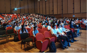 By Ning-Xuan Chen
The 3rd Annual Event of Chinese Young Scholars for OHBM was held on June 11th, during the 2019 OHBM Annual Meeting in Rome. This continued the success from the two previous meetings in Vancouver and Singapore. The theme for this year’s event was “China Roots, Global Impact!” Around 100 young scholars from universities around the world participated. By Roselyne Chauvin
After the success of the first Australian chapter meeting and the announcement of the OHBM publishing platform Aperture in 2018, the OHBM communications committee took the opportunity to meet Michael Breakspear in Rome to get to know more about brain mapping developments in Australia as well as the progress of The OHBM Publishing Initiative Committee (TOPIC). By Ekaterina Dobryakova
Tianzi Jiang is a Director of the Brainnetome at the Institute of Automation the Chinese Academy of Sciences in Beijing, China and a Professor of Neuroimaging and Brainnetome at the Queensland Brain institute of University of Queensland, Australia. The Brainnetome Center attempts to take into consideration the social and environmental effects on the brain of individuals with psychiatric and neurological disorders while examining structural and functional brain characteristics in a multimodal fashion. The Brainnetome atlas, one of the major projects of the Brainnetome Center, currently contains 246 brain regions and allows examination of anatomical connectivity-based parcellation of various brain area. Here we briefly found out about Professor Jiang’s background and his diverse interests centered on the Brainnetome. BY Jessica Turner This year’s OHBM Talairach awardee, Professor Riitta Hari, has had a momentous impact in magnetoencephalography (MEG) research. A professor emerita and Academician of Science and member of the US National Academy of Sciences, she has led the Brain Research Unit of the Low Temperature Laboratory at Aalto University in Finland since 1982. Her work has been critical in understanding how MEG sheds light on brain activity, and how that dynamical activity contributes to cognitive functions including action observation. Here, Jessica Turner found out about Riitta’s background, her current work with artists and the remarkable, undistorted, window into the brain offered by MEG.  Professor Riitta Hari By Ilona Lipp
While there is nothing I would rather research than the brain, I dare say that brain imaging does not always feel like the most rewarding field of science to be in. A single study takes months - more often years - to plan and conduct, the methods can be very expensive and constantly under development, and the results, no matter how interesting, most often just seem like a tiny puzzle piece that (with a lot of luck) will have a (modest) impact in the (distant) future. Coming from this perspective, it was very refreshing for me to talk to somebody whose imaging research is as applied as it can possibly be: Gil Rabinovici. Gil is a professor in neurology, specialized in memory and aging. Using PET imaging with pathology-specific tracers, he does not only investigate mechanisms behind neurodegeneration, but also assesses the clinical applicability and utility of his imaging methods. He recently launched a study on amyloid-PET in more than 18000 people all across the US. Gil will be one of the keynote speakers in Rome and I had the pleasure to find out a bit about him and his research ahead of time. Ilona Lipp (IL): Your research focuses on brain imaging in the context of neurodegeneration and dementia. What are these things called amyloids? |
BLOG HOME
Archives
January 2024
|
 RSS Feed